What Is the Best Imaging for Postoperative Cholesteatoma Surveillance in Adults?
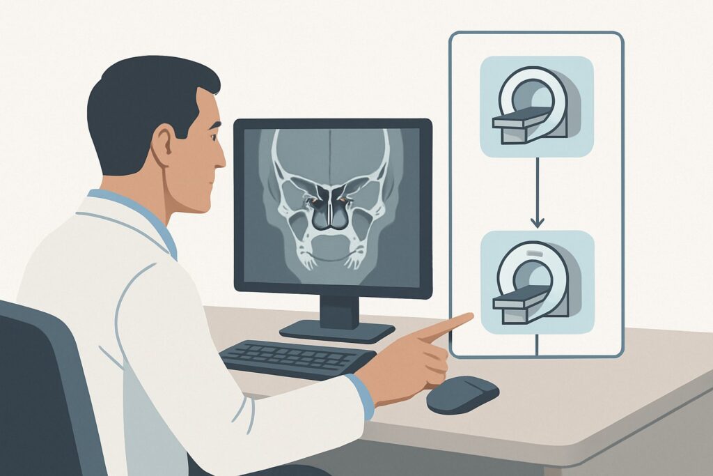
An otolaryngologist is seeing a 48-year-old patient for their one-year follow-up after a canal wall up mastoidectomy for cholesteatoma. The patient feels well, with no new hearing loss, vertigo, or otorrhea. The otoscopic exam, however, is challenging; postoperative changes in the mastoid bowl make it difficult to definitively rule out a small recurrence hidden within the healing tissue. The clinical question is clear: Is there residual or recurrent disease that requires intervention? This article details the American College of Radiology (ACR) evidence-based workflow for this specific surveillance scenario. For this presentation, the ACR Appropriateness Criteria rate MRI head and internal auditory canal without and with IV contrast as Usually Appropriate.
## Who Fits This Clinical Scenario for Postoperative Cholesteatoma Surveillance?
This guidance applies specifically to adults with a history of surgical resection for cholesteatoma who are undergoing routine surveillance imaging. The typical timeframe for the first postoperative scan is 9-12 months after the initial surgery, or later if new, subtle, or equivocal findings arise on clinical examination. The patient is generally asymptomatic or has stable, non-progressive symptoms related to their prior surgery.
This workflow is distinct from several related but different clinical situations. This guidance does not apply to:
- Initial, Preoperative Diagnosis: A patient presenting with signs of chronic otitis media and suspicion of a primary cholesteatoma has different imaging goals, primarily focused on defining the extent of disease for surgical planning. This falls under the Chronic Otitis Media variant.
- Acute Postoperative Complications: A patient presenting days or weeks after surgery with fever, severe pain, facial weakness, or signs of meningitis requires an urgent workup for complications like abscess or sigmoid sinus thrombosis. This is managed under the Complicated Otitis Media pathway.
- Uncomplicated Ear Infections: Patients with symptoms of acute otitis media or otitis externa without a history of cholesteatoma do not typically require advanced imaging.
Correctly identifying the patient as being in a non-acute, surveillance context is critical to selecting the most appropriate and highest-yield imaging study.
## What Diagnoses Are You Working Up with Surveillance Imaging?
In the postoperative setting, the primary goal of imaging is to differentiate benign postoperative changes from recurrent disease. The differential diagnosis is narrow but critical for patient management.
Recurrent or Residual Cholesteatoma
This is the most consequential diagnosis and the primary target of surveillance. Cholesteatoma is a destructive lesion of keratinizing squamous epithelium that can recur if even a small number of cells are left behind. Its propensity to erode the ossicles, scutum, and other temporal bone structures necessitates vigilant monitoring, as recurrence can lead to significant complications, including profound hearing loss, vertigo, facial nerve palsy, and intracranial extension.
Postoperative Granulation Tissue or Scarring
This is the most common benign finding. The middle ear and mastoid heal by forming granulation tissue and, eventually, mature scar tissue. These tissues can appear as soft tissue opacification on imaging and can mimic cholesteatoma on otoscopy, making them a key diagnostic confounder.
Cholesterol Granuloma
Less common, a cholesterol granuloma is an inflammatory, foreign-body giant cell reaction to cholesterol crystals that can occur in any aerated portion of the temporal bone. While benign, it can expand and cause mass effect, and it must be differentiated from cholesteatoma.
Chronic Inflammation
The surgically altered anatomy can sometimes lead to poor aeration and retained fluid, creating an environment for chronic, low-grade inflammation or infection. This can also present as non-specific soft tissue thickening that requires characterization.
## Why Is a Specialized MRI the Recommended Study for Cholesteatoma Surveillance?
The ACR designates MRI head and internal auditory canal without and with IV contrast as Usually Appropriate because of its superior ability to differentiate cholesteatoma from the other soft tissues in the differential diagnosis. The effectiveness of this study hinges on a specific, modern imaging protocol.
The key sequence is Diffusion-Weighted Imaging (DWI), particularly newer, non-echoplanar (non-EPI) techniques. Cholesteatoma, being a dense collection of keratin debris, characteristically demonstrates restricted diffusion, appearing bright on DWI sequences and dark on the corresponding Apparent Diffusion Coefficient (ADC) map. In contrast, granulation tissue, scar, and inflammation do not typically restrict diffusion.
The contrast-enhanced portion of the exam provides complementary information. Recurrent cholesteatoma is an avascular sac of debris and does not enhance. However, surrounding granulation tissue and inflamed mucosa will avidly enhance. This stark difference—a non-enhancing, diffusion-restricting lesion surrounded by enhancing inflammatory tissue—is the classic imaging signature of recurrent cholesteatoma.
Why are other studies rated lower for this specific scenario?
- CT temporal bone without IV contrast: While also rated Usually Appropriate, its role is different. CT provides unparalleled detail of the bony anatomy, including the integrity of the ossicular chain, the facial nerve canal, and the tegmen tympani. However, it cannot reliably distinguish between cholesteatoma, scar tissue, fluid, or granulation tissue, as they all appear as similar-density soft tissue opacification. CT is often considered a complementary study to MRI or a primary alternative if MRI is contraindicated. It involves ionizing radiation (ACR RRL=☢☢☢, 1-10 mSv), a consideration for a condition requiring long-term surveillance.
- Standard MRI head without and with IV contrast: This protocol is rated Usually not appropriate. A routine brain MRI lacks the necessary spatial resolution (thin slices) through the temporal bones and, critically, often omits the specialized non-EPI DWI sequences required to detect small cholesteatomas and avoid susceptibility artifacts that can plague older DWI techniques in this region. Ordering the correct, dedicated protocol is essential for diagnostic accuracy.
The recommended MRI provides the best soft tissue characterization with no ionizing radiation (ACR RRL=O, 0 mSv), making it the ideal first-line modality for surveillance.
## What’s Next After the MRI? Interpreting Results and Planning the Next Steps
The imaging report will guide the subsequent clinical pathway, which should always be determined in collaboration between the radiologist and the treating otolaryngologist.
- Positive for Recurrent Cholesteatoma: If the MRI demonstrates a lesion with the characteristic features of cholesteatoma (restricted diffusion, lack of central enhancement), the next step is typically counseling the patient and planning for revision surgery. The imaging will help define the location and extent of the recurrence, aiding the surgical approach.
- Negative for Recurrence (Benign Findings): If the study shows only stable postoperative changes, enhancing granulation tissue without restricted diffusion, or other benign findings, the patient can typically return to their standard clinical follow-up schedule. This imaging result provides a valuable, reassuring baseline for future comparisons.
- Indeterminate or Equivocal Findings: Sometimes, the imaging findings may be ambiguous. Small lesions or significant imaging artifact can limit diagnostic confidence. In these cases, a multidisciplinary discussion is key. Management options include:
- A shorter interval for follow-up imaging (e.g., in 6 months) to assess for any change.
- Performing a high-resolution non-contrast CT of the temporal bones to correlate soft tissue findings with the underlying bony structures, which may clarify the diagnosis.
- Proceeding to a surgical “second look” procedure, which remains the gold standard for definitive diagnosis when imaging and clinical exams are inconclusive.
## Pitfalls to Avoid (and When to Get Help)
Navigating postoperative surveillance requires attention to detail to avoid common errors that can delay diagnosis or lead to unnecessary procedures.
1. Ordering the Wrong MRI Protocol: The most common pitfall is ordering a “routine MRI of the brain.” This is insufficient. The order must explicitly request MRI of the Head and Internal Auditory Canals for cholesteatoma surveillance, which signals to the radiology department to use a dedicated high-resolution protocol with thin sections and advanced, non-EPI DWI sequences.
2. Misinterpreting DWI Artifacts: Traditional EPI-DWI sequences are prone to susceptibility artifacts at air-bone interfaces, which can mimic or obscure a true cholesteatoma. Interpretation by a neuroradiologist familiar with these pitfalls and the advantages of non-EPI DWI is crucial.
3. Ignoring the Clinical Context: Imaging results should never be interpreted in a vacuum. The patient’s surgical history (e.g., canal wall up vs. down), otoscopic findings, and any new symptoms must be correlated with the imaging report.
4. Delaying Action on Worrisome Findings: If imaging demonstrates extensive bone erosion, particularly concerning for dural or sinus invasion, or if there is any suggestion of intracranial extension, this constitutes a red flag. Urgent consultation with a neurotologist or neurosurgeon is warranted.
## Related ACR Topics and Tools
This article focuses on a single, specific clinical scenario. For a comprehensive overview of imaging for other ear pathologies, from otitis externa to chronic otitis media, please see our parent guide.
- For breadth across all scenarios in Inflammatory Ear Disease, see our parent guide: Inflammatory Ear Disease: ACR Appropriateness Decoded.
To explore adjacent clinical scenarios, review detailed imaging techniques, or discuss radiation dose with patients, the following GigHz resources are available:
- ACR Appropriateness Criteria Lookup — for adjacent scenarios
- Imaging Protocol Library — for technique on the recommended study
- Radiation Dose Calculator — for cumulative dose conversations
Frequently Asked Questions
Why is CT of the temporal bones also rated ‘Usually Appropriate’ for cholesteatoma surveillance?
While MRI is superior for differentiating cholesteatoma from scar tissue, high-resolution CT provides unmatched detail of the bony anatomy. It is excellent for assessing the ossicular chain, facial nerve canal, and subtle bone erosion that might be less conspicuous on MRI. CT is a strong alternative if a patient has a contraindication to MRI (e.g., an incompatible cochlear implant) and is often used as a complementary study to MRI to provide a complete picture of both soft tissue and bone.
How often should surveillance imaging be performed after cholesteatoma surgery?
There is no universal consensus, and practice varies. Many surgeons obtain a baseline scan around one year postoperatively. Subsequent imaging frequency depends on the initial surgical findings, the type of surgery performed (canal wall up vs. down), the results of the baseline scan, and the clinical exam. Asymptomatic patients with a clear initial scan may not need another for several years, while those with equivocal findings may be scanned more frequently.
Can I just order an MRI of the internal auditory canal (IAC) without contrast?
No, this is rated ‘Usually Not Appropriate’ by the ACR for this indication. The non-contrast portion, especially the DWI sequences, is critical for identifying cholesteatoma. However, the post-contrast sequences are essential for differentiating the non-enhancing cholesteatoma from enhancing postoperative granulation tissue or scar. Omitting contrast significantly reduces the diagnostic confidence of the exam.
What is non-EPI DWI and why is it important for cholesteatoma imaging?
Non-EPI DWI (Non-Echo-Planar Imaging Diffusion-Weighted Imaging) is an advanced MRI sequence that is much less susceptible to magnetic field distortion artifacts than conventional DWI. These artifacts are a major problem at the skull base, where bone and air create complex magnetic fields. Non-EPI DWI provides a much cleaner, more reliable image of diffusion, making it far more accurate for detecting small recurrent cholesteatomas in the postoperative mastoid.
Does a normal MRI guarantee there is no recurrent cholesteatoma?
Modern, high-resolution MRI with non-EPI DWI has a very high negative predictive value, meaning a normal scan makes a clinically significant recurrence highly unlikely. However, no imaging test is perfect. Microscopic residual disease would be below the threshold of detection for any imaging modality. Therefore, clinical follow-up with otoscopy remains a critical component of long-term surveillance, even after a negative MRI.
Reviewed by Pouyan Golshani, MD, Interventional Radiologist — May 30, 2026