What Is the Best Imaging for Suspected Differentiated Thyroid Cancer Recurrence?
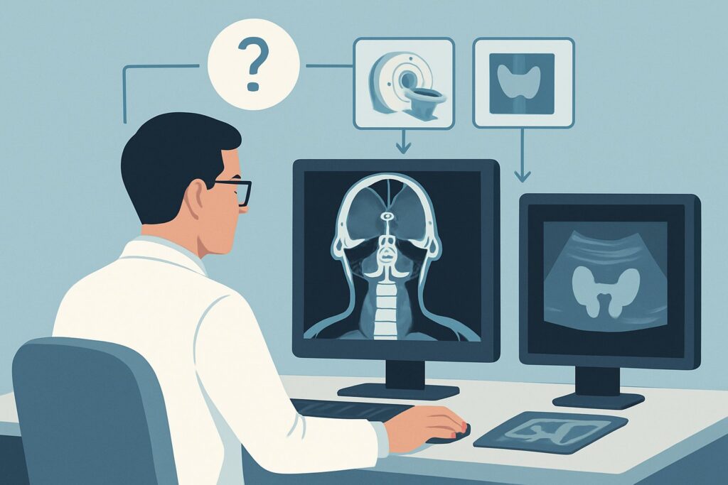
A 58-year-old woman with a history of papillary thyroid cancer, treated seven years ago with a total thyroidectomy and radioactive iodine ablation, presents for her annual follow-up. For the past year, her thyroglobulin (Tg) levels, a key tumor marker, have been slowly but steadily rising despite adequate TSH suppression. Her physical exam is unremarkable, with no palpable masses in the neck. You are now faced with a critical decision: how do you best localize the source of this biochemical recurrence? This article provides a clinical workflow for this exact scenario, detailing why the American College of Radiology (ACR) Appropriateness Criteria rates neck ultrasound as a primary, first-line imaging study. For a patient with suspected recurrence of differentiated thyroid cancer, a `US thyroid` is considered Usually appropriate.
## Who Fits This Clinical Scenario?
This guidance applies specifically to patients with a prior diagnosis of differentiated thyroid cancer (DTC)—which includes papillary, follicular, and Hürthle cell carcinomas—who have completed definitive treatment (typically surgery with or without radioactive iodine). The current presentation is one of suspicion of recurrence, driven by either biochemical evidence (a rising serum thyroglobulin or anti-thyroglobulin antibody level) or a new, concerning clinical finding (such as a palpable nodule in the neck or new hoarseness).
This workflow should be distinguished from several related but distinct clinical situations:
- Initial Nodule Evaluation: This guidance is not for the initial workup of a newly discovered thyroid nodule in a patient without a history of thyroid cancer. That presentation falls under the ACR variant for a palpable thyroid nodule.
- Routine Post-Treatment Surveillance: This is not for asymptomatic, biochemically stable patients undergoing routine, scheduled surveillance imaging in the early post-treatment period. That falls under the variant for early imaging after treatment of differentiated thyroid cancer.
- Non-Differentiated Cancers: This workflow does not apply to medullary or anaplastic thyroid cancers, which have different patterns of spread and require different imaging and management strategies.
The key trigger for this scenario is a change from a stable, disease-free state to one where clinical or laboratory data suggest the cancer has returned.
## What Diagnoses Are You Working Up in This Scenario?
When ordering imaging for suspected DTC recurrence, the primary goal is to locate the disease. The differential diagnosis guides the choice of modality and the interpretation of findings.
The most common consideration is locoregional recurrence in cervical lymph nodes. Differentiated thyroid cancer frequently metastasizes to the lymph nodes of the central neck (level VI, around the trachea and esophagus) and the lateral neck (levels II-V). These nodes may be too small or deep to be palpable, making imaging essential for detection.
Another key possibility is recurrence in the thyroid bed. Even after a total thyroidectomy, microscopic residual thyroid tissue can give rise to a local recurrence. This often appears as a new soft tissue nodule in the surgical bed, which can be difficult to distinguish from post-operative scar tissue without high-resolution imaging and, often, subsequent biopsy.
It is also crucial to consider benign post-surgical changes. Scar tissue (fibrosis), suture granulomas, and benign reactive lymph nodes can all mimic recurrence on imaging or palpation. A high-quality imaging study helps characterize these findings to avoid unnecessary invasive procedures.
Finally, while less common as the sole finding, a rising thyroglobulin level can be the first sign of distant metastases. The most common sites are the lungs and bones. While neck imaging is the appropriate first step to rule out local disease, a negative neck workup in the face of strongly suspicious tumor markers will prompt a search for disease elsewhere.
## Why Is US Thyroid the Recommended Study for This Presentation?
The ACR rates `US thyroid` as Usually appropriate for suspected DTC recurrence because it is the most effective initial tool for evaluating the primary sites of potential disease—the thyroid bed and cervical lymph nodes—without exposing the patient to ionizing radiation.
Ultrasound provides exceptional spatial resolution of the superficial soft tissues of the neck. This allows for detailed characterization of lymph nodes, identifying suspicious features like a rounded (rather than oval) shape, the loss of the normal fatty hilum, the presence of cystic components, or microcalcifications. It is highly sensitive for detecting even small, non-palpable recurrent nodules in the thyroid bed. As a non-invasive study with a radiation level of O 0 mSv, it is ideal for a patient population that may require serial imaging over many years.
Other imaging modalities are rated for specific circumstances:
- CT neck with IV contrast is also rated Usually appropriate. However, it involves a significant radiation dose (☢☢☢ 1-10 mSv) and the use of iodinated contrast. A major pitfall is that iodinated contrast can interfere with the uptake of subsequent diagnostic or therapeutic radioactive iodine (I-131) for weeks or months. Therefore, CT is often reserved for cases where ultrasound is inconclusive, to evaluate for invasion into deeper structures like the airway or major vessels, or to assess bulky disease.
- FDG-PET/CT whole body is rated May be appropriate. This powerful functional imaging study is typically reserved for patients with high biochemical suspicion of recurrence (e.g., significantly elevated Tg) but a negative neck ultrasound and a negative diagnostic I-131 scan. This scenario suggests the recurrence is not avid for iodine but is metabolically active, a phenomenon known as “flip-flop” disease. The radiation dose is substantial (☢☢☢☢ 10-30 mSv).
Once you’ve decided on ultrasound, our protocol guide covers the technique, key anatomical landmarks, and reporting principles: US Thyroid.
## What’s Next After US Thyroid? Downstream Workflow
The results of the neck ultrasound will dictate the subsequent clinical pathway. This is not a one-and-done test but the first step in a diagnostic algorithm.
- If the US is positive: When ultrasound identifies a suspicious nodule in the thyroid bed or an abnormal cervical lymph node, the definitive next step is an ultrasound-guided fine-needle aspiration (FNA). In addition to sending the cytology sample to pathology, it is standard practice to perform a thyroglobulin washout from the needle (FNA-Tg). A high Tg level in the washout fluid is highly specific for metastatic thyroid cancer and can confirm recurrence even if cytology is indeterminate. A confirmed recurrence then leads to discussions about further treatment, which may include surgery, radioactive iodine therapy, or external beam radiation.
- If the US is negative: A negative high-quality neck ultrasound in a patient with low-level or slowly rising Tg may lead to a period of continued observation. However, if biochemical suspicion is high and rising (e.g., Tg > 10 ng/mL), the workup must continue. The next step is often to search for non-iodine-avid or distant disease. This typically involves cross-sectional imaging of the chest (CT) and/or functional imaging with an FDG-PET/CT scan.
- If the US is indeterminate: Sometimes, findings are equivocal, such as a slightly prominent but not overtly malignant-appearing lymph node. In these cases, a short-interval follow-up ultrasound in 3-6 months may be appropriate to assess for stability or change. Alternatively, if clinical suspicion is high enough, one might proceed directly to FNA.
## Pitfalls to Avoid (and When to Get Help)
Navigating the workup for recurrent thyroid cancer requires careful attention to detail to avoid common errors.
- Incomplete Ultrasound Exam: Ensure the sonographer performs a comprehensive bilateral neck survey, including the thyroid bed and all cervical lymph node levels (I-VI). A limited “thyroid only” scan is insufficient.
- Ignoring the Tg/Anti-Tg Trend: A single Tg value is less informative than the trend over time. A rapidly doubling Tg is more concerning than a low, stable level and may warrant a more aggressive workup even with a negative initial ultrasound.
- Premature Use of Iodinated Contrast: Ordering a contrast-enhanced CT scan before considering the potential need for radioactive iodine therapy is a frequent pitfall. The iodine load from the contrast will stun any thyroid tissue, rendering I-131 scans or therapy ineffective for several months.
- Overlooking FNA-Tg Washout: Forgetting to request a thyroglobulin washout during an FNA of a suspicious neck node is a missed opportunity. It can provide a definitive diagnosis when cytology is ambiguous.
If the clinical picture and imaging results are discordant (e.g., very high Tg with negative imaging), it is time to escalate. This typically involves a multidisciplinary tumor board discussion with endocrinology, surgery, radiology, and nuclear medicine to plan the next steps, which often include advanced functional imaging.
## Related ACR Topics and Tools
This article focuses on a single clinical scenario. For a comprehensive overview of imaging for all thyroid-related presentations, from initial nodule workup to goiter and thyrotoxicosis, please see our parent guide.
- For breadth across all scenarios in Thyroid Disease, see our parent guide: Thyroid Disease: ACR Appropriateness Decoded.
To explore other scenarios or understand the technical details of the recommended studies, the following GigHz tools are available:
- ACR Appropriateness Criteria Lookup — for adjacent scenarios
- Imaging Protocol Library — for technique on the recommended study
- Radiation Dose Calculator — for cumulative dose conversations
Frequently Asked Questions
Why is ultrasound preferred over a CT scan if both are rated ‘Usually Appropriate’?
Ultrasound is preferred as the initial study because it provides excellent resolution of the neck’s soft tissues without any ionizing radiation or the need for IV contrast. This avoids the risk of contrast-related complications and, critically, prevents interference from iodinated contrast with potential future radioactive iodine (I-131) scans or therapy. CT is typically reserved for when US is inconclusive or to assess for deep tissue invasion.
What thyroglobulin (Tg) level should trigger an imaging workup for recurrence?
There is no single absolute Tg threshold. The decision is based on the trend and clinical context. A detectable and rising Tg level (e.g., >1-2 ng/mL) in a patient who is post-thyroidectomy and on suppressive therapy is concerning and generally warrants imaging. A rapid doubling time is more alarming than a low, stable value. The presence of anti-thyroglobulin antibodies can interfere with the Tg assay, making the trend of the antibody level itself a potential marker for recurrence.
If the neck ultrasound is negative but my patient’s Tg is 15 ng/mL and rising, what is the next step?
With a high and rising thyroglobulin level but a negative neck ultrasound, the suspicion for disease outside the neck (distant metastases) or in a location poorly visualized by US is high. The next step is typically to pursue functional imaging, most commonly an FDG-PET/CT scan, to identify metabolically active, non-iodine-avid disease. A diagnostic I-131 whole-body scan may also be considered if the patient can be appropriately prepared with TSH stimulation.
Should I order a whole-body iodine scan (I-123 or I-131) as the first test?
While a diagnostic whole-body iodine scan is rated ‘Usually appropriate’ (I-123) or ‘May be appropriate’ (I-131), it is often not the first test performed. It requires the patient to either withdraw from thyroid hormone or receive Thyrogen injections to elevate their TSH, which can be burdensome. Furthermore, its sensitivity for small-volume neck disease can be lower than a high-quality ultrasound. US is simpler, faster, and does not require special patient preparation, making it the preferred initial anatomical imaging test.
Does this guidance apply to medullary thyroid cancer?
No, this guidance is specific to differentiated thyroid cancer (papillary and follicular). Medullary thyroid cancer (MTC) does not typically concentrate iodine, so thyroglobulin is not a useful tumor marker (calcitonin and CEA are used instead), and iodine scans are not used. The imaging workup and surveillance for MTC are different and often involve a combination of neck US, cross-sectional imaging (CT/MRI), and functional imaging like FDG-PET/CT or DOTATATE PET/CT.
Reviewed by Pouyan Golshani, MD, Interventional Radiologist — May 29, 2026