What Is the Best Imaging for Suspected Recurrent Medullary Thyroid Cancer?
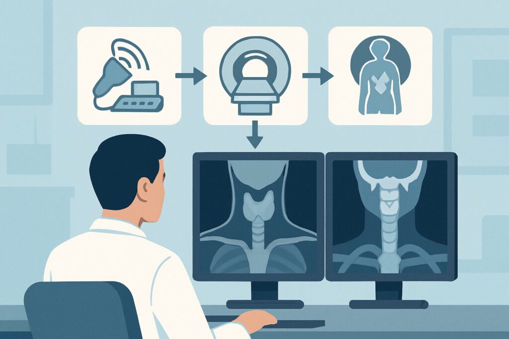
It’s late afternoon, and you receive a message from an endocrinology colleague. A 62-year-old patient, five years post-total thyroidectomy for medullary thyroid cancer (MTC), has shown a steady rise in serum calcitonin on serial labs. The physical exam is unremarkable, but biochemical evidence points toward recurrence. The question is clear: what imaging study should you order first to localize the disease? This article provides a step-by-step workflow for this specific clinical scenario, guiding you through the differential, study rationale, and downstream decisions. For suspected MTC recurrence, the American College of Radiology (ACR) rates neck ultrasound as Usually Appropriate.
Who Fits This Clinical Scenario?
This guidance is for patients with a confirmed history of medullary thyroid cancer who have undergone definitive treatment (typically total thyroidectomy with or without neck dissection) and now present with biochemical evidence of recurrence. The primary trigger is a rising or persistently elevated serum calcitonin level, often accompanied by a rising carcinoembryonic antigen (CEA) level. This workflow is specifically for the post-treatment surveillance phase where the clinical question is localizing recurrent or metastatic disease.
This article does not apply to several similar-sounding situations that require different imaging pathways:
- Initial diagnosis of a thyroid nodule: A patient with a newly discovered palpable thyroid nodule follows a different diagnostic algorithm, starting with imaging to characterize the nodule itself.
- Differentiated thyroid cancer (DTC): Patients with papillary or follicular thyroid cancer have a distinct surveillance protocol. Unlike MTC, DTC often involves radioactive iodine (I-131) scans, as these tumors arise from iodine-avid follicular cells.
- Preoperative staging of MTC: The initial workup for a newly diagnosed MTC involves staging the neck and evaluating for distant disease before surgery, which may involve a broader set of baseline imaging studies.
Correctly identifying your patient’s context—post-treatment MTC with biochemical suspicion of recurrence—is critical to selecting the most effective initial imaging test.
What Diagnoses Are You Working Up in This Scenario?
When calcitonin levels rise after MTC treatment, you are searching for macroscopic disease that may be amenable to further treatment, such as surgery or systemic therapy. The differential is focused on the location and extent of recurrence.
Local-Regional Recurrence
This is the most common pattern of recurrence and the primary target of initial imaging. Disease can reappear in the thyroid bed (the area where the thyroid gland was surgically removed) or, more frequently, in the cervical and upper mediastinal lymph nodes. These recurrences can be small and non-palpable, making high-resolution imaging essential for detection.
Distant Metastases
If local-regional imaging is negative, or if calcitonin levels are very high (e.g., >150-500 pg/mL), the likelihood of distant metastatic disease increases significantly. Medullary thyroid cancer spreads via both lymphatic and hematogenous routes. Common sites for distant metastases include the liver, lungs, and bones. Identifying the site and burden of distant disease is crucial for prognosis and determining the appropriateness of systemic therapies.
Benign Post-Surgical Changes
The post-operative neck can be complex, with scar tissue, surgical clips, and reactive lymph nodes. These changes can sometimes mimic recurrent disease on imaging. A key role of the radiologist is to differentiate these expected post-surgical findings from true tumor recurrence, often by assessing morphology, vascularity, and changes over time compared to prior studies.
Why Is US Thyroid the Recommended Initial Study for This Presentation?
In the setting of suspected MTC recurrence, the ACR designates several studies as Usually Appropriate, but neck ultrasound (US) serves as the logical and effective first step for evaluating the most common site of recurrence: the neck. The rationale is based on its high resolution, lack of radiation, and ability to guide subsequent intervention.
Ultrasound provides exceptional spatial resolution of the soft tissues of the neck, allowing for detailed evaluation of the thyroid bed and the central and lateral cervical lymph node compartments. It can detect subtle abnormalities, such as small, non-palpable nodal metastases, and characterize suspicious features like microcalcifications, cystic changes, and abnormal vascularity. Critically, US is a dynamic examination that can guide fine-needle aspiration (FNA) in real-time if a suspicious lesion is found.
While US is a primary tool, the ACR also rates several other studies as Usually Appropriate, each with a specific role:
- MRI neck without and with IV contrast (0 mSv): An excellent problem-solving tool for the neck, especially when post-surgical changes make ultrasound interpretation difficult or to evaluate for deep extension of disease.
- CT neck with IV contrast (☢☢☢ 1-10 mSv): Provides a comprehensive map of the neck anatomy and is particularly good for assessing larger tumor volumes and their relationship to critical structures.
- CT chest with IV contrast (☢☢☢ 1-10 mSv): This is essential for evaluating the mediastinum and lungs, common sites of MTC metastasis that are beyond the reach of a standard neck ultrasound. It is often performed in conjunction with neck imaging when suspicion for non-local disease is high.
Conversely, some studies are rated lower for a specific reason. For instance, I-131 scan whole body is Usually Not Appropriate. This is a critical distinction: medullary thyroid cancer arises from parafollicular C-cells, which do not concentrate iodine. Therefore, radioactive iodine has no diagnostic or therapeutic role in MTC, unlike in differentiated thyroid cancers. Similarly, DOTATATE PET/CT is also rated Usually Not Appropriate in this initial context, as its utility is not as well-established as other modalities for routine surveillance.
The workflow often begins with the highest-yield, lowest-risk study. Neck US perfectly fits this role, offering detailed anatomical information with no radiation exposure (0 mSv). Once you’ve decided on this initial step, our protocol guide covers the technique, reporting standards, and reading principles. Once you’ve decided on US thyroid, our protocol guide covers the technique, contrast, and reading principles: US Thyroid.
What’s Next After US Thyroid? Downstream Workflow
The results of the neck ultrasound will dictate the next steps in the patient’s management, creating a branching decision tree.
If the Neck US is Positive
If a suspicious lesion (e.g., an abnormal lymph node or soft tissue nodule in the thyroid bed) is identified, the standard next step is a US-guided fine-needle aspiration (FNA). In the context of MTC, this is often coupled with a calcitonin measurement from the needle washout fluid (FNA-Ct). A high FNA-Ct level from the suspicious site confirms metastatic MTC and can guide planning for surgical resection if the disease is localized.
If the Neck US is Negative
A negative high-quality neck ultrasound in a patient with elevated calcitonin is a very common clinical problem. It strongly suggests the recurrence is outside the neck (i.e., distant metastatic disease). The next step depends on the calcitonin level. For moderately elevated levels, cross-sectional imaging of the chest, abdomen, and pelvis is warranted. This typically involves a CT of the chest and a CT or MRI of the abdomen to evaluate the lungs and liver. For very high calcitonin levels or rapidly rising levels, more advanced imaging like FDG-PET/CT (rated May be appropriate) may be considered to search for metabolically active distant disease.
If the Neck US is Indeterminate
When ultrasound findings are equivocal, perhaps due to extensive post-surgical scarring, an MRI neck without and with IV contrast can be a valuable problem-solving tool. Its superior soft tissue contrast may help differentiate scar from recurrent tumor. Alternatively, a short-interval follow-up US in 3-6 months may be appropriate for low-suspicion indeterminate findings.
Pitfalls to Avoid (and When to Get Help)
Navigating the workup for recurrent MTC requires avoiding several common pitfalls to ensure timely and accurate localization of disease.
- Stopping the Search: The most significant error is stopping the workup after a negative neck ultrasound. If biochemical evidence (rising calcitonin) is present, the disease is there somewhere. A negative neck US simply rules out the most common location and mandates a search for distant disease.
- Ordering an Iodine Scan: Never order an I-123 or I-131 scan for a patient with pure medullary thyroid cancer. It provides no useful information and results in unnecessary radiation exposure.
- Underestimating Post-Surgical Anatomy: The operated neck is challenging. Always ensure imaging is interpreted with access to prior studies and surgical reports. Interpretation by a radiologist with head and neck expertise is highly valuable.
If extensive imaging, including neck US and cross-sectional body imaging, remains negative despite significantly elevated or rapidly rising calcitonin, it is time to escalate. This is the definition of “biochemical recurrence with negative conventional imaging,” and management should be discussed in a multidisciplinary tumor board. Referral to a high-volume tertiary care center for consideration of novel imaging agents (e.g., 18F-DOPA PET/CT or 68Ga-somatostatin analogue PET/CT) may be appropriate, often in a clinical trial context.
Related ACR Topics and Tools
This article focuses on a single, specific clinical scenario. For a comprehensive overview of imaging recommendations across all common thyroid-related presentations, from initial nodule workup to thyrotoxicosis, please consult our parent topic hub article. It provides a breadth-first view that complements this deep-dive analysis.
- For breadth across all scenarios in Thyroid Disease, see our parent guide: Thyroid Disease: ACR Appropriateness Decoded.
- To explore other clinical scenarios, use the ACR Appropriateness Criteria Lookup.
- For detailed procedural techniques, see the Imaging Protocol Library.
- To discuss radiation exposure with patients, use the Radiation Dose Calculator.
Frequently Asked Questions
Why is an I-131 radioactive iodine scan not used for medullary thyroid cancer recurrence?
Radioactive iodine (I-131) scans are only effective for cancers that arise from thyroid follicular cells, which have the natural ability to absorb iodine. Medullary thyroid cancer (MTC) originates from the parafollicular C-cells of the thyroid, which do not concentrate iodine. Therefore, an I-131 scan will not detect MTC and is considered ‘Usually Not Appropriate’ for this disease.
My patient’s neck ultrasound is negative, but their calcitonin is over 500 pg/mL. What’s the next step?
A negative neck ultrasound with a significantly elevated calcitonin level (e.g., >150-500 pg/mL) strongly suggests distant metastatic disease. The diagnostic search must be expanded. The next step is typically cross-sectional imaging of the chest, abdomen, and pelvis to evaluate common metastatic sites like the lungs, liver, and bones. This usually involves a CT chest with IV contrast and either a multiphase CT or MRI of the abdomen.
What is the role of FDG-PET/CT in this scenario?
According to the ACR, FDG-PET/CT ‘May be appropriate’ for suspected MTC recurrence. It is generally not a first-line test. Its primary role is in patients with biochemical evidence of recurrence (high calcitonin) but negative conventional imaging (like neck US and CT). In these cases, FDG-PET/CT can sometimes identify metabolically active sites of disease that were not otherwise localized, though its sensitivity can be variable for MTC.
Is there a difference in imaging for sporadic vs. hereditary MTC (as in MEN2 syndromes)?
The imaging principles for localizing recurrent disease are generally the same for both sporadic and hereditary MTC. However, patients with hereditary MTC as part of a Multiple Endocrine Neoplasia (MEN) syndrome require lifelong surveillance for other associated tumors, such as pheochromocytomas and hyperparathyroidism. While the search for recurrent MTC follows this guide, their overall surveillance plan will include additional biochemical and imaging tests tailored to their specific MEN syndrome.
How often should surveillance imaging be performed after MTC treatment if calcitonin levels are stable?
If a patient is in biochemical remission (undetectable or stable low-level calcitonin) after initial treatment, routine surveillance imaging is often not necessary. Surveillance is primarily driven by serial monitoring of serum calcitonin and CEA levels. Imaging is typically initiated only when these tumor markers begin to rise, indicating a high probability of recurrence.
Reviewed by Pouyan Golshani, MD, Interventional Radiologist — May 29, 2026