What Is the Best Imaging for Suspected Wrist Tendon Injury After Normal Radiographs?
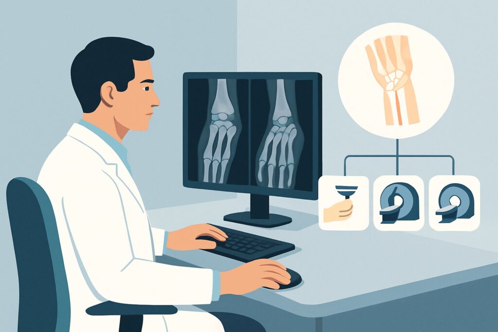
A 48-year-old administrative assistant presents with three months of persistent radial-sided wrist pain, exacerbated by lifting her coffee mug and using a computer mouse. Her initial radiographs were unremarkable. On exam, there is exquisite tenderness over the first dorsal compartment, and a Finkelstein test is positive, raising strong suspicion for de Quervain’s tenosynovitis. You’ve confirmed there’s no obvious fracture or advanced arthritis, but you need to confirm the soft tissue diagnosis and rule out other pathologies before committing to a corticosteroid injection or surgical referral. What is the most appropriate next imaging study? For this specific clinical scenario, the American College of Radiology (ACR) rates Ultrasound (US) of the area of interest as Usually Appropriate.
Who Fits This Clinical Scenario for Suspected Tendon Injury?
This clinical workflow is designed for a specific patient population: adults with chronic hand or wrist pain (lasting several weeks to months) where the initial, standard radiographs are either completely normal or show only nonspecific findings like mild degenerative changes. The crucial element is that your clinical examination points toward a primary tendon or tendon sheath pathology as the source of the pain.
Inclusion criteria for this pathway:
- Adult patient.
- Chronic, non-traumatic hand or wrist pain.
- Focal symptoms that can be localized to a specific tendon or extensor/flexor compartment (e.g., pain over the ECU tendon, snapping with thumb motion).
- Initial radiographs have already been performed and are negative for acute fracture, dislocation, or specific arthritic processes like scapholunate advanced collapse (SLAC wrist).
It is critical to distinguish this presentation from similar, yet distinct, clinical problems that follow different imaging pathways. This guidance does not apply if the primary suspicion is for a different etiology. For instance, if the patient’s symptoms are dominated by numbness and tingling in a classic median nerve distribution, the workup should follow the ACR variant for suspected carpal tunnel syndrome. Similarly, if radiographs showed an old scaphoid fracture, the appropriate next step is to evaluate for nonunion or osteonecrosis, a separate ACR scenario.
What Diagnoses Are You Working Up When Suspecting Tendon Pathology?
When radiographs are unrevealing in a patient with focal chronic wrist pain, the differential diagnosis shifts entirely to the soft tissues. The primary goal of advanced imaging in this context is to visualize the tendons, their sheaths, and adjacent structures to pinpoint the cause of symptoms. The most common considerations include:
Tenosynovitis
This is one of the most common causes of focal wrist pain and involves inflammation and fluid accumulation within the synovial sheath that surrounds a tendon. Classic examples include de Quervain’s tenosynovitis affecting the first dorsal compartment (APL and EPB tendons) and extensor carpi ulnaris (ECU) tenosynovitis on the ulnar side of the wrist. The clinical goal is to confirm the presence and extent of inflammation to guide treatment, such as targeted injections.
Tendinosis (or Tendinopathy)
Unlike the inflammatory nature of tenosynovitis, tendinosis is a degenerative condition characterized by chronic breakdown of the tendon’s collagen fibers. It appears as tendon thickening and internal signal changes on imaging. This is often an overuse injury and can affect tendons like the flexor carpi radialis (FCR). Identifying tendinosis is important as it may respond differently to treatment than acute inflammation.
Partial or Full-Thickness Tendon Tears
While often associated with acute trauma, chronic attritional tears can develop from repetitive microtrauma or impingement. Imaging is crucial to determine the presence, size, and location of a tear, which directly influences whether conservative management is sufficient or if surgical repair is necessary. The ECU tendon is particularly susceptible to tears, often in association with subluxation from its groove.
Ganglion Cysts
These benign, fluid-filled sacs are extremely common around the wrist and can arise from joint capsules or tendon sheaths. While not a tendon pathology themselves, they can cause pain by compressing adjacent nerves or tendons. Imaging can confirm the cystic nature of a palpable lump and define its relationship to nearby structures.
Why Is Ultrasound the Recommended Next Step for Suspected Tendon Injury?
For a patient with normal radiographs and suspected tendon pathology, both Ultrasound (US) of the area of interest and Magnetic Resonance Imaging (MRI) of the area of interest without IV contrast are rated as Usually Appropriate by the ACR. However, ultrasound often serves as the superior first choice due to several key advantages in this specific context.
The primary strength of ultrasound is its ability to perform high-resolution, real-time dynamic imaging. A skilled sonographer can watch the tendon glide as the patient actively moves their wrist or fingers. This is invaluable for diagnosing conditions like ECU tendon subluxation or “snapping” phenomena that are completely invisible on static images like an MRI. Furthermore, the use of color Doppler can instantly visualize hyperemia (increased blood flow), a key sign of active inflammation in tenosynovitis.
From a safety and accessibility standpoint, both US and non-contrast MRI are excellent choices as they involve no ionizing radiation (0 mSv). Ultrasound is typically less expensive, more widely available, and better tolerated by claustrophobic patients than MRI.
Why are other studies rated lower for this scenario?
- MRI without and with IV contrast is rated as May be appropriate. While it provides excellent soft tissue detail, intravenous contrast is rarely necessary to diagnose the common tendon pathologies in this differential. It adds cost and potential risk (e.g., allergic reaction, nephrogenic systemic fibrosis in renal failure) without providing significant additional diagnostic information for this specific question.
- MR Arthrography is rated Usually not appropriate. This invasive procedure, which involves injecting contrast directly into the wrist joint, is designed to evaluate intrinsic structures like the triangular fibrocartilage complex (TFCC) and intercarpal ligaments. It is the wrong tool for assessing extra-articular tendon and tendon sheath pathology.
When ordering, precision is key. A request for “Wrist Ultrasound” is too vague. Instead, specify the exact area of concern and the suspected diagnosis, such as: “Ultrasound of the dorsal radial wrist, please evaluate the first dorsal compartment for de Quervain’s tenosynovitis.” This focuses the examination and yields the most clinically relevant result.
What’s the Next Step After a Wrist Ultrasound? Downstream Workflow
The results of the ultrasound will guide your subsequent management decisions, creating a clear, branching workflow. The goal is to move from diagnosis to definitive treatment efficiently.
If the ultrasound is positive for the suspected pathology:
A definitive finding (e.g., fluid and thickening in the first dorsal compartment consistent with de Quervain’s) confirms the clinical diagnosis. This result provides the confidence to proceed with targeted treatment. For tenosynovitis, this often involves an ultrasound-guided corticosteroid injection, which ensures the medication is delivered precisely into the inflamed tendon sheath, maximizing efficacy and minimizing potential side effects like tendon rupture.
If the ultrasound is negative or inconclusive:
A negative ultrasound in the face of persistent, disabling symptoms should prompt a re-evaluation of the differential diagnosis. The pain may be originating from a deeper structure that ultrasound cannot fully assess, such as bone marrow edema, an occult fracture, or an intrinsic ligament tear. In this situation, the next logical step is to order the other Usually Appropriate study: an MRI of the wrist without IV contrast. MRI provides a more global view of the wrist, including bone, cartilage, and deep ligaments, and may reveal a diagnosis missed on both radiographs and focused ultrasound.
If the ultrasound reveals an unexpected finding:
The study may uncover an alternative diagnosis, such as a ganglion cyst compressing a nerve or an unsuspected partial tendon tear. Each finding has its own management pathway. A symptomatic ganglion may be aspirated under ultrasound guidance, while a significant tendon tear would typically prompt a referral to a hand surgeon for consultation.
Pitfalls to Avoid (and When to Get Help)
Navigating the workup for chronic wrist pain requires avoiding several common pitfalls to ensure an accurate and timely diagnosis.
- Ordering MRI First by Default: While MRI is an excellent test, defaulting to it without considering ultrasound first can miss dynamic pathologies (like tendon subluxation) and is a less cost-effective initial step for superficial tendon issues.
- Vague Imaging Requisitions: A request for “wrist pain” provides no guidance to the radiologist. Always specify the exact location of pain and the suspected clinical diagnosis to ensure a focused, high-yield study.
- Ignoring the Radiographs: Do not skip initial radiographs. They are essential for ruling out bony pathology (fractures, arthritis, tumors) that can mimic soft tissue pain and would lead down a completely different diagnostic path.
- Misinterpreting “Nonspecific Arthritis”: Mild degenerative changes are common and often incidental. Avoid attributing a patient’s focal, severe tendon-related symptoms to mild background osteoarthritis without first ruling out a primary soft tissue cause.
If the diagnosis remains elusive after both a high-quality ultrasound and an MRI, or if the patient’s symptoms are worsening despite initial treatment, it is time to escalate. A consultation with a musculoskeletal radiologist or a referral to a hand surgery specialist is the appropriate next step.
Related ACR Topics and Tools
This article focuses on one specific clinical variant. For a comprehensive overview of imaging for all types of chronic hand and wrist pain, or to explore the rationale for different patient presentations, the following resources are essential.
- For breadth across all scenarios in Chronic Hand and Wrist Pain, see our parent guide: Chronic Hand and Wrist Pain: ACR Appropriateness Decoded.
- ACR Appropriateness Criteria Lookup — Use this tool to find evidence-based recommendations for thousands of clinical scenarios.
- Imaging Protocol Library — Review detailed imaging techniques and protocols for the recommended studies.
- Radiation Dose Calculator — Help patients understand potential radiation exposure from various imaging tests.
Frequently Asked Questions
Why is MRI without contrast also ‘Usually Appropriate’ if ultrasound is preferred?
MRI without contrast is also rated ‘Usually Appropriate’ because it provides excellent overall soft tissue and bone marrow detail. It is a superb alternative if ultrasound is inconclusive, if the patient’s body habitus limits ultrasound visualization, or if there is suspicion of a deeper pathology beyond the reach of an ultrasound probe, such as bone marrow edema or an occult fracture.
Is an ultrasound-guided injection better than a blind injection for tenosynovitis?
Yes, for many locations in the wrist, ultrasound guidance significantly improves the accuracy of a corticosteroid injection. It ensures the medication is delivered directly into the inflamed tendon sheath and avoids inadvertent injection into the tendon itself, which carries a risk of rupture. This precision can lead to better clinical outcomes.
What if the patient’s pain is diffuse and not localized to a specific tendon?
If the pain is diffuse or the clinical exam is non-localizing, this specific workflow may not apply. That presentation fits a different ACR scenario, ‘Adult. Chronic wrist pain. Radiographs normal or remarkable for nonspecific arthritis.’ In that case, MRI without contrast is often preferred over ultrasound as the initial advanced imaging test because it provides a more comprehensive survey of all wrist structures.
Does the presence of ‘nonspecific arthritis’ on the radiograph change the recommendation?
No, not for this scenario. The pathway is designed for cases where radiographs are either normal OR show only nonspecific findings like mild osteoarthritis. As long as the clinical suspicion strongly points to a tendon as the primary pain generator, the presence of mild, likely incidental arthritis does not change the recommendation to proceed with ultrasound or MRI.
When is IV contrast necessary for a wrist MRI?
For the specific indication of suspected tendinopathy or tenosynovitis, IV contrast is rarely needed. However, it becomes essential if there is a clinical concern for other pathologies, such as an infection (abscess), an inflammatory arthritis (like rheumatoid arthritis, to assess for synovitis), or a solid soft-tissue mass or tumor.
Reviewed by Pouyan Golshani, MD, Interventional Radiologist — May 30, 2026