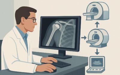
What Is the Best Initial Imaging for an Adult with Acute Traumatic Hip Pain?

A 68-year-old female presents to the emergency department after a ground-level fall onto her left side. She reports immediate, severe pain in her left hip and is unable to bear weight. On examination, the leg is shortened and externally rotated. You need to quickly and efficiently assess for a fracture or dislocation to guide immediate management. This article provides a focused, deep-dive into the American College of Radiology (ACR) guidelines for this exact clinical question: the initial imaging workup for an adult with acute, traumatic hip pain. For this common and critical presentation, the ACR designates `Radiography hip` as Usually Appropriate, establishing it as the clear first-line study.
Who Fits This Clinical Scenario?
This workflow is designed for a specific and frequently encountered patient presentation. The inclusion criteria are straightforward: an adult patient who has experienced a clear traumatic event and now presents with acute hip pain. This typically includes falls (from standing or from a height), motor vehicle collisions, or other direct-impact injuries. The key elements are the acute onset of symptoms immediately following a known trauma.
It is equally important to know when this guidance does not apply. This workflow is not intended for:
- Patients with suspected occult fracture: If the initial radiographs are negative or indeterminate but your clinical suspicion for a fracture remains high (e.g., the patient cannot bear weight), you have moved into a different clinical scenario. The next imaging step is addressed in a separate ACR variant.
- Patients with known, positive fractures on radiographs: If the initial x-ray clearly shows a fracture, the next step is often preoperative planning, which may or may not require advanced imaging like CT. This is a distinct downstream workflow.
- Patients with atraumatic hip pain: If the pain began without any injury, the differential diagnosis is much broader (including avascular necrosis, arthritis, infection, or tumor), and the imaging algorithm is different.
Correctly identifying your patient’s scenario ensures you order the right initial test and avoid unnecessary or low-yield studies.
What Diagnoses Are You Working Up in This Scenario?
In the setting of acute trauma, the differential diagnosis is focused on urgent orthopedic injuries that require prompt diagnosis and management. The initial imaging choice is tailored to rapidly confirm or exclude these specific conditions.
Femoral Neck or Intertrochanteric Fracture: This is the primary concern, especially in older adults with osteoporosis after a fall. These fractures can disrupt blood supply to the femoral head (particularly intracapsular femoral neck fractures) and almost always require surgical intervention. Radiographs are the cornerstone of diagnosis due to their high accuracy for displaced fractures.
Hip Dislocation: A less common but equally urgent diagnosis, typically resulting from high-energy trauma like a car accident. Posterior dislocations are most common. Prompt reduction is critical to prevent complications like avascular necrosis of the femoral head or sciatic nerve injury. Radiographs are excellent for identifying the dislocation and any associated fractures.
Pelvic Ring or Acetabular Fractures: The trauma may have been significant enough to fracture the pelvis or the acetabulum (the “socket” of the hip joint). While often visible on standard hip radiographs, dedicated pelvic views or CT may be needed for full characterization, especially if surgery is contemplated. The initial hip radiograph serves as a crucial screening tool.
Greater or Lesser Trochanter Avulsion Fractures: These can occur from forceful muscle contractions during trauma, particularly in younger, athletic individuals. While less severe than femoral neck fractures, they cause significant pain and functional limitation and are typically well-visualized on radiographs.
Why Is Hip Radiography the Recommended Initial Study for This Presentation?
The American College of Radiology designates `Radiography hip` as Usually Appropriate for the initial evaluation of an adult with acute, traumatic hip pain. This recommendation is based on the modality’s high diagnostic yield for the most critical injuries, its speed, wide availability, and favorable safety profile.
Radiographs are highly sensitive and specific for detecting displaced fractures and dislocations, which are the most common and urgent considerations in this scenario. A standard series, typically including an anteroposterior (AP) view of the pelvis and both AP and lateral views of the symptomatic hip, provides a comprehensive initial assessment of the femoral head, neck, trochanteric regions, and acetabulum.
In contrast, other powerful imaging modalities are rated lower for this initial step:
- Computed Tomography (CT) of the hip is rated Usually Not Appropriate as a first-line test. While excellent for characterizing complex fractures found on an x-ray, it is not the recommended initial study. Using CT first exposes the patient to significantly more radiation (☢☢☢ 1-10 mSv, same as radiography but for a much more detailed study) without adding diagnostic value over radiographs for the primary question of “is there a fracture or dislocation?” Its role is in preoperative planning or in cases of high-energy trauma where pelvic ring injury is strongly suspected.
- Magnetic Resonance Imaging (MRI) of the hip is also rated Usually Not Appropriate for initial imaging. MRI is the most sensitive test for detecting occult (radiographically invisible) fractures, bone marrow edema, and soft-tissue injuries. However, it is slower, more expensive, and less available in an emergency setting. Its primary role is as a second-line test when initial radiographs are negative but clinical suspicion for a fracture remains high.
The choice of radiography balances diagnostic utility with resource stewardship and patient safety. It rapidly answers the most pressing clinical question, using a low dose of ionizing radiation (ACR Relative Radiation Level ☢☢☢ 1-10 mSv) to guide immediate patient care.
What’s Next After Hip Radiography? Downstream Workflow
The results of the initial hip radiograph directly guide the subsequent clinical pathway. The decision tree branches based on whether the findings are positive, negative, or indeterminate.
If the radiograph is POSITIVE for a fracture or dislocation: The next step is orthopedic consultation for management, which is typically surgical for displaced hip fractures or requires urgent reduction for dislocations. The orthopedic surgeon may order a CT scan, particularly for complex fracture patterns involving the acetabulum or for preoperative planning to better define fracture fragments. This moves the patient into the “Radiographs positive for hip fracture. Next imaging study” scenario.
If the radiograph is NEGATIVE but clinical suspicion remains high: This is a critical juncture. If a patient has significant pain, focal tenderness over the femoral neck, and an inability to bear weight despite normal-appearing x-rays, an occult hip fracture is a strong possibility. In this case, the workup is not complete. The next appropriate step is to proceed to a more sensitive imaging study. This places the patient into the ACR scenario, “Suspect fracture. Radiographs negative or indeterminate. Next imaging study,” for which MRI without contrast is the most appropriate test.
If the radiograph is INDETERMINATE: Sometimes, findings can be ambiguous due to patient positioning, body habitus, or overlying structures. If a fracture cannot be confidently excluded, the patient should be managed as if a fracture is present (e.g., non-weight-bearing status) and further imaging should be pursued, typically with MRI or, if MRI is contraindicated, CT or a nuclear medicine bone scan.
Pitfalls to Avoid (and When to Get Help)
In the acute setting, several common pitfalls can delay diagnosis or lead to misinterpretation. Be mindful of the following:
- Stopping the workup after a negative x-ray in a high-suspicion patient: The “hip fracture” that is most commonly missed is the non-displaced femoral neck fracture in an elderly patient who cannot bear weight. A negative radiograph does not rule out a fracture in this context.
- Inadequate imaging technique: A true lateral view of the hip is essential for evaluating femoral neck alignment and subtle fractures. An incomplete or poorly positioned study can obscure pathology. Do not hesitate to ask for repeat views if the initial images are suboptimal.
- Overlooking associated injuries: In high-energy trauma, always examine the entire pelvis on the AP pelvic view, not just the symptomatic hip. Associated pubic rami fractures or sacroiliac joint widening can be subtle but important clues to a more significant pelvic ring injury.
If the clinical picture and radiographic findings are discordant, or if you suspect a complex pelvic injury, early consultation with orthopedics and/or a radiologist is crucial to ensure the correct downstream imaging and management plan are initiated promptly.
Related ACR Topics and Tools
This article covers one specific variant within the broader topic of Acute Hip Pain. For a comprehensive overview of all related scenarios, from atraumatic pain to postoperative follow-up, please consult our parent guide. The following GigHz tools can also support your clinical decision-making:
- For breadth across all scenarios in Acute Hip Pain, see our parent guide: Acute Hip Pain: ACR Appropriateness Decoded.
- ACR Appropriateness Criteria Lookup — To explore imaging recommendations for adjacent or alternative clinical scenarios.
- Imaging Protocol Library — For detailed technical specifications on how imaging studies like hip radiography and CT are performed.
- Radiation Dose Calculator — To help in discussions with patients about cumulative radiation exposure from medical imaging.
Frequently Asked Questions
Why not just order a CT scan first for everyone with traumatic hip pain?
While CT is excellent for detailed bone anatomy, it is rated ‘Usually Not Appropriate’ as the initial test by the ACR for this scenario. A standard radiograph is faster, more widely available, uses less radiation, and is highly effective at answering the primary clinical question: is there a displaced fracture or dislocation? CT is reserved for cases where the radiograph is positive and more detail is needed for surgical planning, or in select high-energy trauma cases.
What if the patient is pregnant? Is a hip radiograph still the first choice?
Yes, radiography is generally still the initial imaging modality of choice, even in pregnancy. The radiation dose to the fetus from a properly shielded hip and pelvis radiograph is very low and well below the threshold known to cause harm. The risk of missing a significant, mobility-altering fracture far outweighs the minimal radiation risk. MRI is an alternative if there is a very high clinical concern and a desire to avoid all ionizing radiation, but it is often not necessary as the first step.
Does this guidance apply to chronic hip pain after an old injury?
No. This workflow is specifically for acute pain immediately following a new traumatic event. Chronic or subacute pain, even if it started after a remote injury, falls under a different clinical category. The differential for chronic pain is much broader, including post-traumatic arthritis, avascular necrosis, or non-union, and the imaging workup would be different.
The radiograph was negative, but the patient can’t walk. What is the single best next test?
When initial radiographs are negative in a patient with a high clinical suspicion for fracture (e.g., inability to bear weight, focal bony tenderness), you have moved to the ‘suspected occult fracture’ scenario. The ACR designates MRI without IV contrast as the most appropriate next study. MRI is extremely sensitive for detecting bone marrow edema associated with a non-displaced fracture that is invisible on a plain x-ray.
Is ultrasound useful for traumatic hip pain?
For the initial evaluation of a suspected fracture or dislocation, ultrasound is rated ‘Usually Not Appropriate.’ It cannot adequately visualize the femoral neck or deep bony structures. Ultrasound’s role in hip imaging is primarily for evaluating soft tissues, such as looking for a joint effusion, tendinopathy, or muscle tears, which are secondary concerns in the initial trauma workup focused on bone injury.
Reviewed by Pouyan Golshani, MD, Interventional Radiologist — May 30, 2026