What Imaging Is Best for Foot Trauma When Ottawa Rules Are Negative but Forefoot Pain Persists?
A 35-year-old patient presents to your urgent care clinic after dropping a heavy box on their right foot an hour ago. They can bear weight and walk, albeit with a limp. On examination, you find no tenderness over the posterior edge of the medial or lateral malleolus, nor at the base of the fifth metatarsal or the navicular bone. The Ottawa Ankle and Foot Rules are negative. However, the patient has exquisite point tenderness and swelling over the shaft of the third metatarsal. You suspect a fracture in an area not covered by the Ottawa rules. What is the appropriate initial imaging study to order in this common clinical scenario?
According to the American College of Radiology (ACR) Appropriateness Criteria, the correct initial study is Radiography foot, which is rated Usually appropriate. This article provides a detailed workflow for this specific presentation, outlining the differential diagnosis, the rationale for radiography over other modalities, and the next steps based on imaging results.
Who Fits This Clinical Scenario for Acute Foot Trauma?
This guidance applies to a specific and frequently encountered patient presentation: an adult or a child over 5 years of age with acute foot trauma where the Ottawa Ankle and Foot Rules can be fully evaluated and are negative. The key feature of this scenario is that clinical suspicion for injury remains high, but the pain is localized to an anatomic area not addressed by the Ottawa rules. This typically includes the forefoot (metatarsals, metatarsal-phalangeal joints), the toes (phalanges), or associated soft tissue structures like tendons.
It is critical to distinguish this situation from several similar-but-distinct clinical scenarios that follow different imaging pathways:
- Positive Ottawa Rules: If the patient has pain in the malleolar or midfoot zones AND either bone tenderness at specific points (posterior edge of malleoli, navicular, base of the fifth metatarsal) OR an inability to bear weight, the Ottawa rules are positive. This indicates a higher likelihood of a clinically significant ankle or midfoot fracture and mandates imaging based on those findings.
- Inability to Evaluate Ottawa Rules: If the patient cannot cooperate with the examination due to factors like intoxication, altered mental status, significant distracting injuries, or diminished sensation, the Ottawa rules are not applicable. These patients typically require radiography regardless of the physical exam.
- High Suspicion for Lisfranc Injury: If the mechanism of injury (e.g., axial load on a plantarflexed foot) or specific physical exam findings (e.g., plantar ecchymosis, instability with forefoot manipulation) suggest a Lisfranc ligamentous complex injury, the workup is more urgent and may require weight-bearing radiographs or advanced imaging from the outset.
This article is exclusively for the patient with negative Ottawa rules but persistent, localized pain elsewhere in the foot.
What Diagnoses Are You Working Up When Ottawa Rules Are Negative?
When the Ottawa rules are negative, you have effectively ruled out a high probability of a clinically significant fracture in the ankle and midfoot. The diagnostic focus shifts to the forefoot and toes, where the patient’s symptoms are localized. The differential diagnosis in this scenario includes several common injuries.
The most frequent and important diagnosis to confirm or exclude is a metatarsal fracture. These often result from direct blows, crush injuries, or twisting forces. While some metatarsal fractures are stable and heal well with conservative care, others, like fractures of the fifth metatarsal base (e.g., a Jones fracture) or significantly displaced fractures, require orthopedic consultation and potentially surgical intervention.
A phalangeal (toe) fracture is another primary consideration, especially with a history of direct impact or a “stubbing” injury. While most toe fractures are managed non-operatively with buddy taping and supportive footwear, imaging is valuable to assess for intra-articular extension, significant displacement, or angulation that might warrant reduction.
Less commonly, the injury may involve the joints or soft tissues. A metatarsophalangeal (MTP) joint sprain or dislocation, such as “turf toe” affecting the first MTP joint, can result from hyperextension. While primarily a soft-tissue injury, radiographs are essential to rule out an associated avulsion fracture. Finally, a severe soft tissue contusion or hematoma can mimic a fracture with significant pain and swelling. In this case, imaging serves to confidently exclude a bony injury, allowing for appropriate symptomatic treatment.
Why Is Foot Radiography the Recommended First Step in This Scenario?
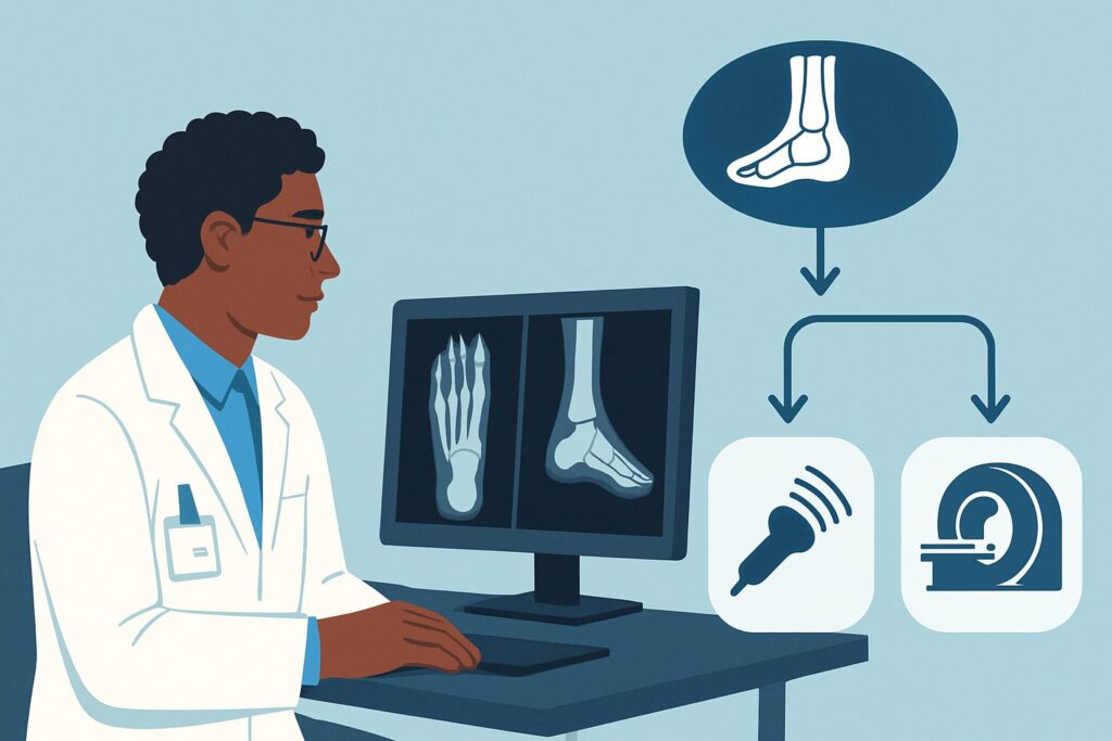
For a patient with suspected forefoot or toe pathology after acute trauma, standard foot radiography is the cornerstone of initial evaluation. The ACR rates both Radiography foot and Radiography foot with weightbearing as Usually appropriate for this specific clinical scenario.
The rationale is straightforward: radiography is an excellent first-line tool for identifying or excluding the most common and clinically important injuries in this context—metatarsal and phalangeal fractures. It is fast, widely accessible, inexpensive, and provides clear visualization of bony anatomy. The radiation dose is extremely low, with an effective dose of less than 0.1 mSv for adults and less than 0.03 mSv for children, making it a safe choice for both populations. If there is any concern for subtle ligamentous injury or instability, obtaining weight-bearing views can be particularly helpful and does not significantly increase the radiation exposure.
Alternative imaging modalities are generally not indicated for the initial workup and receive lower appropriateness ratings from the ACR for valid reasons:
- CT foot without IV contrast is rated May be appropriate (Disagreement). While CT provides exquisite bony detail and is superior for detecting occult fractures or defining complex fracture patterns, it is not necessary as a first step. It involves a higher radiation dose, particularly for pediatric patients (☢☢ 0.03-0.3 mSv), and greater cost. The “Disagreement” among the panel highlights its role as a problem-solving tool for cases where radiographs are negative but clinical suspicion for a fracture remains very high.
- US foot and MRI foot without IV contrast are both rated Usually not appropriate. Ultrasound is not a reliable tool for a comprehensive fracture survey of the foot. MRI is highly sensitive for soft tissue injuries (tendons, ligaments) and bone marrow edema, but it is expensive, less available, and unnecessary for the primary goal of ruling out a simple forefoot fracture. These modalities are reserved for downstream evaluation if a soft tissue injury is suspected after a fracture has been excluded.
What Is the Downstream Workflow After a Foot Radiograph?
The results of the initial foot radiograph directly guide the subsequent clinical management and decision-making process. The workflow typically branches into one of three paths.
If the radiograph is positive for a fracture: The next step depends on the specific fracture’s location, displacement, and angulation. For a simple, non-displaced phalangeal fracture, treatment often involves buddy taping and a hard-soled shoe. For a non-displaced metatarsal shaft fracture, a walking boot and protected weight-bearing may be sufficient. However, if the radiograph reveals a more complex injury—such as a displaced fracture, an intra-articular fracture, or a high-risk fracture like a Jones fracture at the fifth metatarsal base—prompt consultation with an orthopedic specialist is warranted to discuss potential reduction or surgical fixation.
If the radiograph is negative: A negative X-ray in the setting of persistent, severe point tenderness requires careful clinical judgment. If suspicion for an occult fracture remains high, the patient can be treated conservatively (e.g., with a walking boot and instructions for non-weight-bearing) with a plan for close follow-up in 7-10 days. At that point, repeat radiographs may show a previously invisible fracture line. Alternatively, if a definitive diagnosis is needed sooner, proceeding to CT foot without IV contrast (May be appropriate) can identify occult fractures. If the clinical picture is more suggestive of a ligamentous or tendon injury, an MRI may be considered.
If the radiograph is negative and clinical suspicion is low: If the pain is manageable and the exam is reassuring despite the initial trauma, the likely diagnosis is a soft tissue contusion or sprain. The patient can be managed symptomatically with rest, ice, compression, elevation (RICE), and analgesics, with instructions to return if symptoms worsen or fail to improve.
Pitfalls to Avoid (and When to Get Help)
When evaluating a patient with acute foot trauma and negative Ottawa rules, several common pitfalls can lead to missed diagnoses or improper management.
- Misapplying the Ottawa Rules: A frequent error is assuming that negative Ottawa rules rule out all foot fractures. Remember, the rules are only validated for the midfoot and ankle. This entire clinical scenario is built on recognizing that the rules do not apply to the forefoot or toes.
- Overlooking Sesamoid Injuries: Fractures of the two small sesamoid bones beneath the great toe’s MTP joint can be a source of significant pain and disability. These can be subtle and are sometimes missed on standard views; dedicated sesamoid views may be necessary if tenderness is localized there.
- Failing to Consider Weight-Bearing Views: For injuries where ligamentous stability is a concern, non-weight-bearing films may appear normal. If the mechanism or exam suggests instability, obtaining weight-bearing views is crucial and is also rated Usually appropriate.
- Dismissing a Clinically Suspicious Negative Radiograph: A single negative radiograph does not definitively rule out a fracture, especially in the early stages. If a patient has severe, focal bony tenderness that persists, trust your clinical exam and arrange for appropriate follow-up or advanced imaging. If you are uncertain about a subtle finding on a radiograph, escalate by obtaining a formal radiology read or consulting with an orthopedic specialist.
Related ACR Topics and Tools
For a comprehensive overview of all clinical variants related to acute foot trauma, as well as detailed information on other imaging topics, the following resources are available.
- For breadth across all scenarios in Acute Trauma to the Foot, see our parent guide: Acute Trauma to the Foot: ACR Appropriateness Decoded.
- To explore other clinical scenarios and their corresponding ACR ratings, use the ACR Appropriateness Criteria Lookup.
- For detailed technical specifications of imaging studies, consult the Imaging Protocol Library.
- To discuss radiation exposure with patients, the Radiation Dose Calculator can help contextualize the dose from medical imaging.
Frequently Asked Questions
Why not just order an MRI initially to see everything, including soft tissues and occult fractures?
While MRI is excellent for soft tissue and bone marrow edema, it is rated ‘Usually not appropriate’ as an initial study in this scenario. Radiography is faster, cheaper, more widely available, and highly effective for the primary question: is there a fracture in the forefoot or toes? An MRI is considered overkill for this initial evaluation and is best reserved for cases where radiographs are negative but a significant soft tissue (ligament/tendon) injury is suspected.
If the Ottawa rules are negative, is it ever acceptable to not order any imaging?
Yes. The decision to image is based on clinical suspicion. If a patient has negative Ottawa rules and only minimal, diffuse tenderness in the forefoot that is improving, it is reasonable to diagnose a soft tissue contusion and manage conservatively without imaging. This ACR scenario applies specifically when there is persistent, focal tenderness or a high clinical suspicion for a fracture despite the negative Ottawa rules.
Does this guidance apply to a suspected stress fracture from overuse?
No, this guidance is for acute trauma. The workup for a suspected stress fracture is different. Initial radiographs for a stress fracture are often negative. If suspicion remains high, the next appropriate step is often an MRI, which is highly sensitive for detecting the bone marrow edema associated with a stress injury.
What is the difference between a standard foot radiograph and one with weight-bearing?
A standard foot radiograph is taken with the patient lying down (non-weight-bearing). A weight-bearing radiograph is taken while the patient is standing on the affected foot. This can reveal subtle instability or widening between bones (e.g., in a Lisfranc injury) that may not be apparent on non-weight-bearing views. For a suspected simple metatarsal or toe fracture from a direct blow, weight-bearing views are often not necessary, but they are rated equally as ‘Usually appropriate’ and can add value if ligamentous stability is a concern.
Is CT really ‘May be appropriate’ if it has more radiation than an X-ray?
Yes, the ‘May be appropriate (Disagreement)’ rating reflects its specific role as a second-line, problem-solving tool. It should not be used first. However, if a radiograph is negative and you have a very high suspicion for a complex or occult fracture that would change management (e.g., an intra-articular fracture), the diagnostic benefit of CT can outweigh the additional radiation risk. The panel’s disagreement indicates this is not a routine choice but a reasonable one in select circumstances.
Reviewed by Pouyan Golshani, MD, Interventional Radiologist — June 27, 2026