What Is the Best Initial Imaging for Stepwise Cognitive Decline or Post-Stroke Dementia?

A 74-year-old man with a history of hypertension and a transient ischemic attack (TIA) six months ago is brought to your clinic by his daughter. She reports a noticeable, stepwise decline in his cognitive function since the TIA. He was sharp before, but now struggles with multi-step tasks and has become more forgetful. His decline isn’t a smooth, gradual slide; it seems to worsen in distinct steps. You suspect a vascular component to his cognitive impairment. The central question is what initial imaging study will best clarify the diagnosis and guide management. This article provides a detailed clinical workflow for this specific scenario, explaining why the American College of Radiology (ACR) finds an MRI of the head without IV contrast to be Usually Appropriate.
Who Fits This Clinical Scenario for Suspected Vascular Dementia?
This guidance applies specifically to adult patients presenting with cognitive impairment where a vascular etiology is the leading suspicion. The key clinical features that place a patient in this category are a history of a recent stroke or a documented “stepwise” or “stuttering” decline in cognitive function. This pattern, often characterized by periods of stability followed by abrupt deterioration, is the clinical hallmark of vascular dementia and distinguishes it from the more insidious, gradual decline seen in other neurodegenerative conditions.
This workflow is not intended for patients with:
- A primary, insidious memory deficit without a clear vascular event. This presentation is more typical of Alzheimer disease and routes to a different ACR Appropriateness Criteria variant.
- Prominent early behavioral changes or progressive aphasia. If personality changes, disinhibition, or language difficulties are the dominant initial symptoms, the workup should be oriented toward suspected frontotemporal dementia.
- Mild cognitive impairment (MCI) not yet meeting the criteria for dementia. While vascular disease can contribute to MCI, the pre-dementia workup follows a distinct pathway.
Correctly identifying the patient’s clinical pattern is crucial for selecting the most informative initial imaging study.
What Diagnoses Are You Working Up with Imaging in This Scenario?
When ordering imaging for a patient with a suspected vascular component to their dementia, you are primarily investigating the presence, location, and burden of cerebrovascular disease. The differential diagnosis is centered on conditions where vascular injury is the primary or a significant contributing driver of cognitive loss.
The most common consideration is Vascular Dementia (VaD). This is not a single disease but a syndrome resulting from various forms of cerebrovascular pathology. Imaging seeks to identify the underlying cause, which could be multi-infarct dementia from multiple cortical or subcortical strokes, or subcortical ischemic vascular dementia (often called Binswanger’s disease) resulting from extensive chronic small-vessel disease affecting the deep white matter.
A highly prevalent and important diagnosis is Mixed Dementia, where both vascular pathology and Alzheimer disease pathology coexist. Vascular insults can lower the threshold for the clinical expression of Alzheimer’s, and the two pathologies often interact synergistically. Imaging can reveal evidence of both—such as chronic infarcts alongside medial temporal lobe atrophy—which has significant prognostic and management implications.
Less common but consequential diagnoses include Strategic Infarct Dementia, where a single, small infarct in a functionally critical brain region (like the thalamus or angular gyrus) causes a disproportionately severe cognitive deficit. Imaging is essential to pinpoint such a lesion. Another key consideration is Cerebral Amyloid Angiopathy (CAA), a condition characterized by amyloid deposition in cerebral blood vessels, leading to lobar microhemorrhages and cognitive decline. It often co-occurs with Alzheimer disease but has distinct imaging features.
Why Is MRI of the Head Without Contrast the Recommended Initial Study?
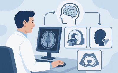
The ACR designates an MRI of the head without IV contrast as Usually Appropriate for this clinical scenario because of its superior ability to characterize the full spectrum of cerebrovascular pathology. MRI provides detailed anatomical information that is critical for differentiating between the potential diagnoses and assessing the overall burden of vascular disease.
MRI is highly sensitive for detecting key findings that are often subtle or invisible on other modalities. Standard MRI sequences can clearly depict:
- Chronic Infarcts: Both large territorial infarcts and small, deep lacunar infarcts are readily identified.
- White Matter Hyperintensities: T2-weighted and FLAIR sequences are exceptionally sensitive for showing the extent of chronic small-vessel ischemic change, a cornerstone of the diagnosis.
- Microhemorrhages: Susceptibility-weighted imaging (SWI) or gradient-recalled echo (GRE) sequences are essential for detecting tiny hemorrhages, which are the hallmark of cerebral amyloid angiopathy.
- Atrophy Patterns: MRI can assess for global or focal volume loss, helping to identify co-existing neurodegenerative patterns, such as the medial temporal lobe atrophy characteristic of Alzheimer disease.
In contrast, alternative studies are rated lower for specific reasons. A CT of the head without IV contrast is rated May be appropriate (Disagreement). While it can identify large chronic strokes and is useful when MRI is contraindicated or unavailable, it has poor sensitivity for small lacunar infarcts, microhemorrhages, and the true extent of white matter disease. An MRI of the head without and with IV contrast is rated Usually not appropriate because, in the routine initial evaluation for vascular dementia, gadolinium-based contrast adds little diagnostic information and introduces unnecessary risk and cost. Contrast is typically reserved for cases where other pathologies like tumor, infection, or inflammation are suspected.
This recommended study involves no ionizing radiation (adult RRL=O 0 mSv).
What’s Next After MRI of the Head Without Contrast? Downstream Workflow
The results of the non-contrast head MRI will guide the subsequent clinical pathway, focusing on risk factor modification, symptomatic treatment, and patient counseling.
- If the study is positive for significant vascular pathology (e.g., multiple infarcts, extensive white matter disease): The diagnosis of vascular dementia or mixed dementia is supported. The next steps are aggressive management of vascular risk factors: strict blood pressure control, statin therapy, diabetes management, and antiplatelet therapy as indicated for secondary stroke prevention. The focus shifts from diagnosis to mitigating further vascular damage.
- If the study is negative or shows only minimal, age-expected changes: A primary vascular etiology becomes less likely. The workup should pivot to reconsider other causes of dementia. This may involve re-evaluating for a primary neurodegenerative process like Alzheimer disease (which may require different imaging, such as amyloid PET, in atypical cases) or exploring less common dementia mimics.
- If the study is indeterminate or shows unexpected findings (e.g., features suggestive of inflammation or a mass): Further characterization is needed. This is a scenario where a follow-up MRI with IV contrast might be appropriate, or a referral to a neurologist or neuroradiologist for consultation on the next steps is warranted. If the initial MRI suggests significant stenosis of the large intracranial or cervical vessels, a dedicated vascular imaging study like MRA or CTA may be considered.
The MRI report provides the anatomical basis for a comprehensive management plan tailored to the patient’s specific pathology.
Pitfalls to Avoid (and When to Get Help)
In the workup of suspected vascular dementia, several common pitfalls can delay diagnosis or lead to suboptimal management.
- Accepting a “negative” CT as ruling out vascular pathology: A non-contrast head CT can miss the majority of the evidence for small-vessel vascular dementia. If clinical suspicion is high, an MRI is necessary.
- Over-attributing all cognitive decline to “old strokes”: The presence of chronic infarcts doesn’t automatically equal vascular dementia. The clinical history, pattern of decline, and imaging findings must be correlated to exclude a co-existing and potentially treatable condition.
- Ignoring microhemorrhages: The presence and pattern of microhemorrhages on SWI/GRE sequences are critical. A report noting numerous lobar microbleeds should raise suspicion for CAA, which has implications for the safety of anticoagulants and antiplatelet agents.
If the clinical picture and imaging findings are discordant, or if the MRI reveals complex or unexpected pathology, consultation with a neurologist or cognitive specialist is the appropriate next step.
Related ACR Topics and Tools
This article focuses on a single clinical scenario. For a comprehensive overview of imaging for all dementia presentations, from Alzheimer disease to frontotemporal dementia, please see our parent guide. For additional resources to help select and understand imaging studies, the following tools are available.
- For breadth across all scenarios in Dementia, see our parent guide: Dementia: ACR Appropriateness Decoded.
- ACR Appropriateness Criteria Lookup — for adjacent scenarios
- Imaging Protocol Library — for technique on the recommended study
- Radiation Dose Calculator — for cumulative dose conversations
Frequently Asked Questions
Why is a non-contrast MRI preferred over one with contrast for this initial workup?
For the initial evaluation of suspected vascular dementia, IV contrast is rated ‘Usually not appropriate’ by the ACR. The key diagnostic features—chronic infarcts, white matter disease, microhemorrhages, and atrophy—are all well-visualized on standard non-contrast sequences (like FLAIR, DWI, and SWI/GRE). Contrast does not improve the detection of these findings and is reserved for cases where other pathologies like a brain tumor, infection, or active inflammation are suspected based on atypical clinical or imaging features.
If my patient has a pacemaker and cannot get an MRI, is a CT scan a good enough substitute?
A non-contrast head CT is rated ‘May be appropriate (Disagreement)’ and is the best alternative if MRI is absolutely contraindicated. However, it is a significant compromise. CT can identify large chronic strokes but is insensitive to small lacunar infarcts, the full extent of white matter ischemic disease, and microhemorrhages. A ‘normal’ CT scan does not rule out vascular dementia. The report should be interpreted with the understanding that significant small-vessel pathology may be present but not visible.
Should I order an MRA of the head or neck at the same time as the initial brain MRI?
MRA of the head or neck is rated ‘May be appropriate.’ It is not a required part of the initial dementia workup but can be considered if there is a clinical suspicion of large-vessel stenosis contributing to the patient’s cognitive decline or if the patient has had recent ischemic events suggesting an embolic source. It is often ordered as a separate consideration based on the broader stroke-prevention workup rather than the dementia evaluation itself.
What specific MRI sequences are most important for this diagnosis?
A comprehensive brain MRI protocol for dementia should include: 1) T2-weighted FLAIR to assess white matter hyperintensities, 2) Diffusion-Weighted Imaging (DWI) to identify acute or subacute infarcts, 3) Susceptibility-Weighted Imaging (SWI) or Gradient-Recalled Echo (GRE) to detect chronic microhemorrhages, and 4) T1-weighted sequences to evaluate brain volume and atrophy patterns, particularly of the medial temporal lobes.
Does functional imaging like FDG-PET or SPECT have a role in this scenario?
For the initial workup of suspected vascular dementia, functional imaging like FDG-PET or SPECT is rated ‘Usually not appropriate’ by the ACR. These studies assess metabolic activity and perfusion but are less specific for vascular pathology than structural MRI. Their primary role in dementia imaging is for differentiating neurodegenerative disorders, such as distinguishing Alzheimer disease from frontotemporal dementia, when the diagnosis is unclear after structural imaging.
Reviewed by Pouyan Golshani, MD, Interventional Radiologist — May 29, 2026