What Is the Best Initial Imaging for Suspected Breast Implant-Associated Malignancy?

A 55-year-old patient with textured silicone implants placed over a decade ago presents to your clinic. For the past month, she has noticed progressive, painless swelling of her left breast, which now feels firm and larger than the right. On examination, there is a palpable fluid wave and no discrete, dominant mass. You are concerned about a delayed seroma and the possibility of a breast implant-associated malignancy. The immediate clinical question is which imaging study to order first to safely and effectively begin the workup. According to the American College of Radiology (ACR) Appropriateness Criteria, for this initial evaluation, breast ultrasound is rated Usually Appropriate.
Who Fits This Clinical Scenario for Suspected Implant-Associated Malignancy?
This clinical workflow applies to any adult female or transfeminine patient with any type of breast implant (saline or silicone; smooth or textured) who presents with new signs or symptoms concerning for an implant-associated malignancy. The key feature is a change from their baseline post-operative state, often occurring years after implant placement.
Inclusion criteria for this pathway include:
- A new, spontaneous, and persistent peri-implant fluid collection (seroma).
- A new palpable mass adjacent to the implant or in the axilla.
- Capsular contracture that is new, rapidly progressing, or associated with pain or a mass.
- Overlying skin changes such as rash, erythema, or ulceration without a clear infectious cause.
It is crucial to distinguish this presentation from other common implant-related issues that follow different diagnostic pathways. This guidance does not apply to patients presenting with asymptomatic concerns for implant integrity or those with a clear history of trauma leading to suspected implant rupture. For instance, a patient presenting for routine, asymptomatic screening for silicone implant rupture or a patient with sudden deflation of a saline implant would be evaluated under different ACR criteria variants.
What Diagnoses Are You Working Up in This Scenario?
When a patient presents with symptoms concerning for an implant-associated malignancy, the imaging workup is designed to investigate a specific, though narrow, differential diagnosis. The primary goal is to identify features that warrant tissue or fluid sampling.
Breast Implant-Associated Anaplastic Large Cell Lymphoma (BIA-ALCL) is the principal diagnosis of concern. This is a rare T-cell lymphoma that is not a cancer of the breast tissue itself but arises from the scar tissue capsule that forms around the implant. The most common presentation, seen in the majority of cases, is a delayed, spontaneous seroma. Less commonly, it can present as a solid mass on the implant capsule. It has a stronger association with textured-surface implants.
Benign Late Seroma is a more common cause of peri-implant fluid collection and represents a diagnosis of exclusion. While benign, its clinical presentation is indistinguishable from that of BIA-ALCL. Therefore, any significant late seroma must be sampled to definitively rule out malignancy.
Primary Breast Cancer must always be considered. The presence of implants does not protect against the development of typical breast adenocarcinomas. A new mass or axillary lymphadenopathy could represent a primary breast cancer that is unrelated to the implant itself. Imaging must be capable of evaluating both the implant capsule and the surrounding breast parenchyma.
Implant Rupture with Granuloma (Siliconoma) can occur with silicone implant rupture, where leaked silicone incites a significant inflammatory response, forming a firm mass that can mimic a malignancy. While this is a benign process, it can be a clinical and imaging confounder.
Squamous Cell Carcinoma (SCC) and other rare carcinomas arising from the implant capsule have also been described. These are exceedingly rare but are part of the differential for a solid mass associated with the implant capsule.
Why Is Breast Ultrasound the Recommended First Study for This Presentation?
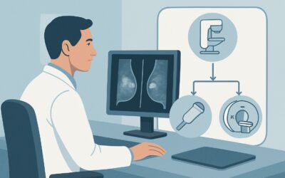
For the initial evaluation of suspected breast implant-associated malignancy, the ACR designates breast ultrasound as Usually Appropriate. This recommendation is based on its high utility for identifying the key findings in this scenario, its safety profile, and its accessibility.
Ultrasound is exceptionally effective at characterizing peri-implant fluid collections, the hallmark presentation of BIA-ALCL. It can accurately quantify the volume of the seroma and assess for any associated abnormalities, such as thickening of the implant capsule or an adjacent solid mass. Most importantly, ultrasound provides real-time guidance for fine-needle aspiration of the fluid, which is the essential next diagnostic step. The procedure is safe, minimally invasive, and allows for the collection of an adequate sample for cytology, flow cytometry, and cell block with immunohistochemistry (IHC) for CD30 and Anaplastic Lymphoma Kinase (ALK) markers.
Furthermore, ultrasound has no ionizing radiation (relative radiation level of 0 mSv) and does not require intravenous contrast. It is also excellent for evaluating the axillary and other regional lymph nodes for suspicious adenopathy.
Other imaging modalities are rated differently for this specific initial workup:
- Mammography or Digital Breast Tomosynthesis are rated May be appropriate. While important for general breast cancer screening, they are less sensitive for evaluating peri-implant fluid and subtle capsular abnormalities. Implants can obscure visualization of the capsule, and the compression required can be uncomfortable for a patient with a tense seroma. They also involve a low dose of ionizing radiation (0.1-1 mSv).
- MRI breast without and with IV contrast is also rated Usually Appropriate but is typically reserved as a second-line or problem-solving tool. While highly sensitive for detecting capsular masses and assessing the extent of disease, it is more costly, less accessible, and often not necessary if ultrasound clearly identifies a seroma for aspiration. An MRI breast without IV contrast, however, is rated Usually not appropriate because contrast is essential for characterizing masses and inflammation; a non-contrast study is primarily for evaluating implant integrity, which is not the primary clinical question here.
Therefore, ultrasound serves as the ideal first-line modality, directly answering the most pressing question—is there a fluid collection or mass that needs sampling?—and guiding the subsequent intervention.
What’s Next After Breast Ultrasound? A Downstream Workflow
The results of the initial breast ultrasound will dictate the subsequent management steps in a clear, branching pathway. The goal is to move efficiently toward a definitive diagnosis.
If ultrasound confirms a significant peri-implant fluid collection (typically >50 cc):
The immediate next step is an ultrasound-guided aspiration of the seroma. It is critical to send the aspirate for a specific panel of tests: cytology to look for atypical cells, flow cytometry, and cell block for CD30 and ALK immunohistochemistry. A positive CD30 stain with negative ALK is characteristic of BIA-ALCL.
If ultrasound identifies a solid mass associated with the implant capsule or an abnormal lymph node:
The next step is an ultrasound-guided core needle biopsy of the solid component. This provides a tissue sample for histopathologic evaluation, which is necessary to diagnose BIA-ALCL presenting as a mass or to identify another type of malignancy like SCC or primary breast cancer.
If ultrasound is negative or equivocal but clinical suspicion remains high:
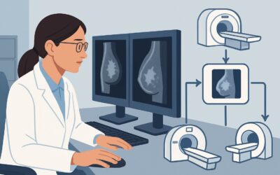
If the patient has persistent symptoms but the ultrasound is unrevealing, this is the primary indication to proceed with a breast MRI with and without IV contrast. MRI offers superior soft tissue contrast and can detect subtle capsular enhancement, small masses, or adenopathy that may be missed on ultrasound.
If BIA-ALCL is diagnosed:
A confirmed diagnosis triggers a multidisciplinary team evaluation involving surgical oncology, medical oncology, and plastic surgery. Standard management involves complete surgical excision of the mass, the implant, and the entire surrounding fibrous capsule (total capsulectomy). Further systemic staging with a PET/CT scan is typically performed to assess for disease beyond the breast.
Pitfalls to Avoid (and When to Get Help)
Navigating the workup for suspected implant-associated malignancy requires careful attention to detail to avoid common missteps. A primary pitfall is failing to aspirate a new, spontaneous peri-implant seroma, instead attributing it to benign causes without confirmation. Any such fluid collection requires sampling. Another error is sending the aspirated fluid only for routine cytology, without the specific request for CD30 and ALK immunohistochemistry, which are essential for diagnosing BIA-ALCL. Finally, do not order a non-contrast breast MRI to evaluate a mass; contrast is mandatory for tissue characterization. If the initial ultrasound is negative but your clinical suspicion is high due to persistent, unexplained symptoms, escalate the workup to a contrast-enhanced breast MRI and consider referral to a breast specialist.
Related ACR Topics and Tools
This article focuses on a single clinical scenario. For a comprehensive overview of all related presentations, from asymptomatic screening to suspected rupture, please consult our parent guide. For additional resources on imaging selection, protocols, and radiation safety, the following GigHz tools are available.
- For breadth across all scenarios in Breast Implant Evaluation, see our parent guide: Breast Implant Evaluation: ACR Appropriateness Decoded.
- For adjacent scenarios not covered here, use the ACR Appropriateness Criteria Lookup.
- For detailed procedural techniques, explore the Imaging Protocol Library.
- For discussions about cumulative exposure, reference the Radiation Dose Calculator.
Frequently Asked Questions
Does the type of breast implant (saline vs. silicone) change the initial imaging recommendation?
No, for the initial workup of a suspected implant-associated malignancy, the recommendation for breast ultrasound as the first-line study remains the same regardless of whether the implant is saline or silicone-filled. The primary concern is evaluating the capsule and peri-implant space for fluid or masses, which ultrasound does effectively for both types.
If BIA-ALCL is suspected, should both breasts be imaged?
Yes, a bilateral breast ultrasound, including evaluation of both axillae, is recommended. Although the patient’s symptoms may be unilateral, BIA-ALCL can, in rare instances, be bilateral. A complete bilateral examination provides a comprehensive baseline and ensures no contralateral abnormalities are missed.
How soon after implant placement can BIA-ALCL occur?
BIA-ALCL is typically a delayed complication. The median time from implant placement to diagnosis is approximately 8 to 10 years. It is very uncommon to see it develop within the first few years post-implantation.
Is a PET/CT scan appropriate for the initial diagnosis?
No, PET/CT is not recommended for the initial diagnosis. Its role is in staging the disease *after* a tissue or fluid diagnosis of BIA-ALCL has already been confirmed. The initial workup should focus on ultrasound to identify a target for sampling, followed by MRI if needed.
If a patient has a known implant rupture, does that increase their risk for BIA-ALCL?
Currently, there is no established evidence to suggest that implant rupture (either saline or silicone) directly increases the risk of developing BIA-ALCL. The primary risk factor identified to date is the surface texture of the implant, with textured implants carrying a significantly higher risk than smooth-surfaced implants.
Reviewed by Pouyan Golshani, MD, Interventional Radiologist — May 30, 2026