What Is the Next Imaging Study for a Suspected Stress Fracture After Negative X-Rays?
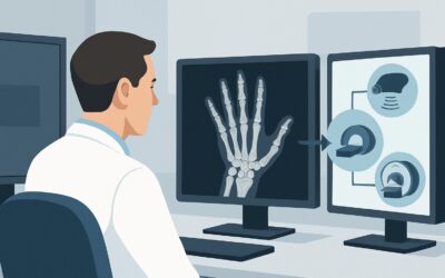
A 24-year-old marathon runner presents with 3 weeks of worsening, localized anterior shin pain. She recently increased her training intensity and now has point tenderness over the mid-tibia. The pain is sharp with running and subsides with rest. You suspect a tibial stress fracture, but the initial radiographs you ordered are unremarkable. This common clinical crossroads requires a definitive next step to confirm the diagnosis, guide treatment, and prevent a complete fracture. This article details the American College of Radiology (ACR) guided workflow for an adult with a suspected stress fracture and negative or indeterminate initial radiographs. For this specific scenario, the ACR rates an MRI of the area of interest without IV contrast as Usually Appropriate.
Who Fits This Clinical Scenario for a Suspected Stress Fracture?
This workflow is designed for a specific patient population: an adult with a clinical suspicion for a stress fracture (either a fatigue fracture from abnormal stress on normal bone or an insufficiency fracture from normal stress on abnormal bone) located anywhere except the vertebrae. The crucial element of this scenario is that initial imaging with radiographs has already been performed and was either negative or indeterminate, failing to confirm the diagnosis.
This guidance does not apply to several similar-but-distinct clinical situations, which have their own recommended pathways:
- Initial Imaging: This article is for the second step in the workup. If you have not yet ordered any imaging, please refer to the ACR variant for initial imaging in a suspected stress fracture.
- Pregnant Patients: The workup for a suspected pelvic or hip stress fracture in a pregnant patient involves different considerations to avoid ionizing radiation to the fetus and follows a separate ACR guideline.
- High Risk for Fracture Completion: Patients with known severe osteoporosis or concerning radiographic signs (like the “dreaded black line” in an anterior tibial stress fracture) may require a different or more urgent imaging approach to assess the risk of progression to a complete, displaced fracture.
Correctly identifying that your patient fits this scenario—symptoms persisting despite negative initial X-rays—is key to ordering the most effective next study.
What Diagnoses Are You Working Up When Radiographs Are Negative?
When focal bone pain persists despite normal radiographs, the differential diagnosis broadens. The goal of advanced imaging is to distinguish the most likely cause from less common but important mimics.
Stress Fracture: This remains the primary diagnosis. Radiographs are notoriously insensitive in the early stages of a stress injury, often remaining normal for several weeks. The underlying pathophysiology—an imbalance between bone resorption and formation due to repetitive stress—initially manifests as microfractures and bone marrow edema, which are invisible on X-ray. Advanced imaging is needed to detect these early changes.
Medial Tibial Stress Syndrome (MTSS) or “Shin Splints”: Particularly relevant for lower leg pain, MTSS is a traction-induced periostitis, an inflammation of the bone’s outer lining (periosteum) where muscles attach. Its symptoms can closely mimic a tibial stress fracture, but the management and prognosis differ. Imaging can precisely differentiate between periosteal edema (MTSS) and a true stress fracture with marrow edema and a potential fracture line.
Tendon or Ligament Injury: Chronic insertional tendinopathy or ligamentous strain can cause focal pain near a bony prominence, mimicking a stress injury. For example, pain near the navicular bone in the foot could be a high-risk navicular stress fracture or posterior tibial tendinopathy.
Occult Neoplasm: Though less common, a primary bone tumor (like an osteoid osteoma) or a metastatic lesion can present with focal, activity-related pain. This is a critical “can’t-miss” diagnosis, especially in older patients or those with a known primary malignancy. Advanced imaging is essential for evaluating for an underlying lesion.
Osteomyelitis: A bone infection can also cause localized pain and be radiographically occult in its early stages. While the clinical context is often different (e.g., fever, overlying soft tissue infection), it remains in the differential for unexplained focal bone pain.
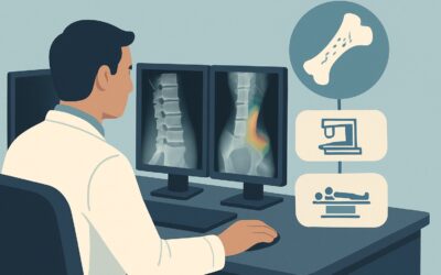
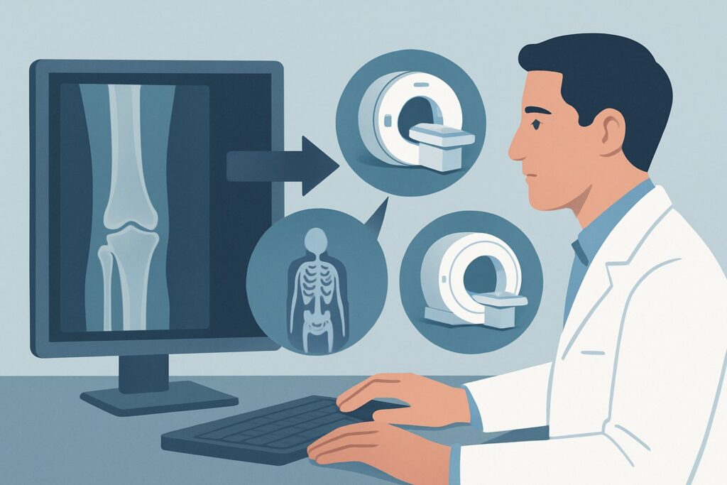
Why Is MRI Without Contrast the Recommended Next Study for a Suspected Stress Fracture?
When radiographs are unrevealing, Magnetic Resonance Imaging (MRI) of the area of interest without IV contrast is rated Usually Appropriate and is the diagnostic test of choice. Its high sensitivity for early bone stress changes and lack of ionizing radiation make it the superior option in this scenario.
The primary strength of MRI is its ability to detect bone marrow edema, the earliest sign of a stress reaction. This appears as a high signal on fluid-sensitive sequences (like STIR or T2-fat-suppressed images) long before a cortical fracture line becomes visible on any other imaging modality. This allows for a definitive diagnosis weeks earlier than repeat radiographs. Furthermore, MRI can grade the severity of the injury (e.g., using the Fredericson classification), which helps guide activity modification and predict time to recovery. It also provides excellent evaluation of the surrounding soft tissues, allowing for confident diagnosis of mimics like tendinopathy or muscle injury.
Other imaging modalities are rated lower for specific reasons:
- Repeat Radiography in 10-14 days is rated May be appropriate. While inexpensive, this approach inherently delays diagnosis. If periosteal reaction or a fracture line appears, the diagnosis is confirmed. However, a negative repeat X-ray still does not definitively rule out a stress injury, often forcing the clinician to order advanced imaging anyway, but now two weeks later.
- Bone Scan (Scintigraphy) is rated May be appropriate. A technetium-99m bone scan is highly sensitive and will detect the increased bone turnover of a stress fracture early. However, it is not specific; infection, tumor, and other inflammatory conditions can also appear as “hot spots.” It also exposes the patient to a moderate dose of ionizing radiation (☢☢☢ 1-10 mSv). MRI provides superior anatomic detail to characterize the finding without radiation.
- CT without IV contrast is also rated May be appropriate. CT is excellent for delineating a subtle cortical fracture line but is less sensitive than MRI for detecting the early marrow edema of a stress reaction. It is a good problem-solving tool in select cases but is not the recommended primary advanced imaging modality due to its use of ionizing radiation and lower sensitivity for early-stage injury.
An MRI without IV contrast is sufficient. The addition of gadolinium-based contrast is rated Usually not appropriate as it does not add diagnostic information for a typical stress fracture workup, while increasing cost and carrying a small risk of adverse reactions.
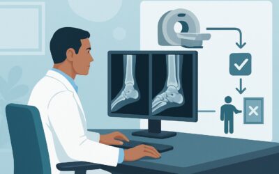
What’s Next After MRI? Downstream Workflow
The results of the MRI will guide your next clinical steps. The decision tree is typically straightforward.
If the MRI is positive for a stress fracture: The diagnosis is confirmed. Management focuses on relative rest, activity modification, and addressing underlying causes (e.g., training errors, biomechanics, nutritional deficiencies). The grade of the stress injury on MRI can help determine the duration of non-weight-bearing or modified activity. For high-risk fractures (e.g., femoral neck, anterior tibia, navicular), an orthopedic consultation is warranted, as these may require more aggressive management, including potential surgery, to prevent complications like complete fracture or avascular necrosis.
If the MRI is negative for a stress fracture: A stress fracture is effectively ruled out. The high-resolution images will often reveal the alternative cause of pain, such as medial tibial stress syndrome, tendinopathy, or a muscle tear. Management should be redirected to treat this specific diagnosis. If the MRI is completely normal and the patient’s symptoms persist, further clinical re-evaluation is needed to consider non-musculoskeletal causes of pain.
If the MRI shows an unexpected finding (e.g., tumor or infection): The finding dictates the next step. A suspected bone tumor requires urgent referral to an orthopedic oncologist for biopsy and further management. Suspected osteomyelitis requires consultation with infectious disease specialists and orthopedic surgery for potential debridement and targeted antibiotic therapy.
Pitfalls to Avoid (and When to Get Help)
Navigating this workflow requires avoiding a few common pitfalls to ensure timely and accurate diagnosis.
- Delaying Advanced Imaging: Relying too long on the “wait-and-see” approach with repeat radiographs can delay diagnosis, prolonging the patient’s pain and potentially increasing the risk of the stress fracture progressing to a complete fracture.
- Ignoring High-Risk Locations: Not all stress fractures are equal. Be highly suspicious of pain in the femoral neck, patella, anterior tibial cortex, medial malleolus, talus, and navicular. These are considered high-risk locations, and a confirmed diagnosis on MRI should prompt a low threshold for orthopedic consultation.
- Ordering the Wrong MRI: Ordering an MRI “with and without contrast” is unnecessary for this indication. This adds cost and potential risk with no benefit. Be specific on your order: “MRI [body part] without contrast to evaluate for stress fracture.”
If a high-risk stress fracture is identified or if the MRI reveals a concerning lesion like a tumor, escalate care immediately by consulting with an orthopedic surgeon.
Related ACR Topics and Tools
For a comprehensive overview of all clinical variants related to this topic, from initial imaging to special populations, please see the parent guide. For tools to help with ordering, protocoling, and discussing imaging with patients, the following resources are available.
- For breadth across all scenarios in Stress (Fatigue/Insufficiency) Fracture, Including Sacrum, Excluding Other Vertebrae, see our parent guide: Stress (Fatigue/Insufficiency) Fracture, Including Sacrum, Excluding Other Vertebrae: ACR Appropriateness Decoded.
- ACR Appropriateness Criteria Lookup — for adjacent scenarios
- Imaging Protocol Library — for technique on the recommended study
- Radiation Dose Calculator — for cumulative dose conversations
Frequently Asked Questions
Why not just get a CT scan instead of an MRI? It’s often faster.
While CT is faster, it is less sensitive than MRI for detecting early-stage stress fractures. CT excels at showing fine cortical bone detail and can identify a subtle fracture line, but it cannot visualize bone marrow edema, which is the earliest sign of a stress injury. Because MRI can detect these changes weeks earlier and involves no ionizing radiation, it is the preferred next step after negative radiographs.
Is a bone scan a reasonable alternative if MRI is not available?
A bone scan is rated ‘May be appropriate’ and can be a reasonable alternative if MRI is contraindicated or unavailable. It is very sensitive for detecting areas of high bone turnover, so it will be positive early in the course of a stress fracture. However, its main drawback is a lack of specificity—many other conditions, like infection or tumor, can also cause a positive bone scan. It also involves a moderate dose of radiation.
Do I need to order the MRI with contrast?
No. For the evaluation of a suspected stress fracture, IV contrast is not necessary and is rated ‘Usually not appropriate’ by the ACR. The key diagnostic findings, such as bone marrow edema and a potential fracture line, are clearly visualized on non-contrast sequences. Adding contrast increases cost and adds a small risk of adverse reaction without providing additional diagnostic value for this specific indication.
What if the patient’s pain is diffuse and not well-localized? Should I still order an MRI?
If the pain is diffuse, an MRI of a specific area may not be the best initial advanced study. In cases of poorly localized pain where the differential is broad, a whole-body bone scan may be more useful to identify a potential ‘hot spot’ that can then be further evaluated with a targeted MRI. However, if there is a point of maximal tenderness, even with some surrounding diffuse pain, an MRI of that specific area remains the most appropriate test.
My patient is an adolescent athlete, not an adult. Does this same guidance apply?
While the ACR scenario specifies an adult patient, the diagnostic principles are very similar for adolescents. MRI without contrast is still the imaging modality of choice for suspected stress fractures with negative radiographs in pediatric and adolescent patients, particularly due to its lack of ionizing radiation. However, the differential diagnosis in children can be broader, including physeal (growth plate) injuries, apophysitis, and different primary bone tumors, which must be considered.
Reviewed by Pouyan Golshani, MD, Interventional Radiologist — May 30, 2026