What Is the Right First Imaging Study for Chronic Elbow Pain? ACR Workflow
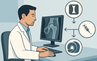
A 58-year-old patient presents to your clinic with several months of aching pain in their right elbow, worse with activity. There was no specific injury, and the exam reveals diffuse tenderness without instability, locking, or focal neurologic signs. As you open the electronic medical record to place an order, you face a common clinical question: what is the most appropriate initial imaging study to work up undifferentiated chronic elbow pain? This article provides a detailed workflow for this specific scenario, explaining why the American College of Radiology (ACR) designates standard radiography as the clear first step. For this presentation, **Radiography elbow** is rated *Usually Appropriate*.
Who Fits This Clinical Scenario for Initial Elbow Imaging?
This guidance applies to adult patients presenting with chronic elbow pain—pain lasting for weeks to months—where the clinical picture is not yet specific. The key inclusion criterion is the chronicity of symptoms without a clear acute traumatic event and in the absence of dominant, localizing signs that point to a specific pathology. The patient’s primary complaint is pain, which may be intermittent or constant, and is often exacerbated by use of the arm.
It is critical to distinguish this general presentation from more specific scenarios that follow different diagnostic pathways. This workflow is not appropriate if:
- The patient reports prominent mechanical symptoms. If the chief complaint includes locking, catching, or a distinct loss of motion, the workup shifts to evaluating for intra-articular bodies or other structural derangements.
- There is high suspicion for a specific soft-tissue injury refractory to treatment. If the exam points strongly to lateral or medial epicondylalgia (tennis or golfer’s elbow) or a tendon tear that has failed conservative management, the imaging strategy changes to directly evaluate those structures.
- Focal neurologic symptoms are present. If the patient describes paresthesias, weakness, or pain radiating in a specific nerve distribution (e.g., ulnar nerve), the workup must prioritize evaluation of potential nerve entrapment.
This article focuses exclusively on the initial imaging choice for undifferentiated chronic elbow pain, where the goal is to efficiently screen for common underlying causes.
What Diagnoses Are You Working Up with Initial Elbow Radiographs?
When ordering the initial study for chronic elbow pain, the goal is to evaluate for common and consequential pathologies that are readily visible on radiographs. The differential diagnosis at this stage is broad but anchored by conditions affecting the bone and joint space.
Osteoarthritis (OA): This is one of the most common causes of chronic joint pain, particularly in middle-aged and older adults or those with a history of manual labor or prior trauma. Radiographs are highly effective at identifying the classic signs of OA, including joint space narrowing (particularly of the radiocapitellar and ulnohumeral joints), osteophyte formation, and subchondral sclerosis.
Inflammatory Arthritis: Conditions such as rheumatoid arthritis or psoriatic arthritis can manifest in the elbow. While symptoms are often polyarticular, the elbow can be a primary site of pain. Radiographs can reveal characteristic findings like symmetric joint space loss, periarticular osteopenia, and bony erosions that would prompt a rheumatologic workup.
Intra-articular Loose Bodies: Fragments of bone or cartilage can break off and float within the joint, causing pain and sometimes mechanical symptoms. If these bodies are calcified or ossified, they are often visible on standard radiographs. Their presence can confirm the need for orthopedic referral, potentially for arthroscopy.
Occult Bony Abnormality: Less commonly, chronic pain may be the result of a subtle, non-displaced fracture that was missed acutely, a stress fracture from repetitive overuse, or an underlying bony lesion. Radiographs serve as an essential first-line screening tool to detect changes in bone density, cortical disruptions, or other structural abnormalities that would require further investigation.
Why Are Elbow Radiographs the ‘Usually Appropriate’ First Step?
The ACR designates **Radiography elbow** as *Usually Appropriate* for the initial imaging of chronic elbow pain because it is a high-value test that directly and efficiently assesses for the most common bony and articular causes. It provides a crucial baseline evaluation with minimal radiation exposure and cost, guiding subsequent management or more advanced imaging if necessary.
The strength of radiography in this scenario lies in its ability to confirm or exclude the key osseous pathologies in the differential diagnosis. It is highly sensitive and specific for detecting degenerative changes, articular calcifications, and most fractures or bony lesions. Answering these primary questions first prevents unnecessary and costly advanced imaging.
In contrast, more advanced modalities are rated lower for this *initial*, undifferentiated presentation:
- MRI elbow without IV contrast is rated *Usually not appropriate* as a first step. While MRI provides exquisite detail of soft tissues like tendons, ligaments, and nerves, ordering it before radiographs is an inefficient use of resources. Many causes of chronic elbow pain are bony, and if radiographs identify significant osteoarthritis, an expensive MRI may not change the initial non-operative management plan. MRI is reserved for when radiographs are negative but clinical suspicion for a soft-tissue abnormality remains high.
- US elbow is also rated *Usually not appropriate* for this initial, generalized workup. Ultrasound is a powerful tool for focused clinical questions, such as evaluating the common extensor tendon in suspected lateral epicondylitis. However, for non-specific pain, it provides a limited field of view and cannot adequately assess the entire joint for osseous pathology. Its utility is in targeted evaluation, not broad screening.
From a safety perspective, elbow radiography involves a very low radiation dose (relative radiation level ☢ <0.1 mSv), making it a safe starting point. This compares favorably to CT, which delivers a higher dose (☢☢ 0.1-1mSv) and is also rated *Usually not appropriate* for this initial workup due to its primary role in detailed bone assessment after an abnormality is already suspected.
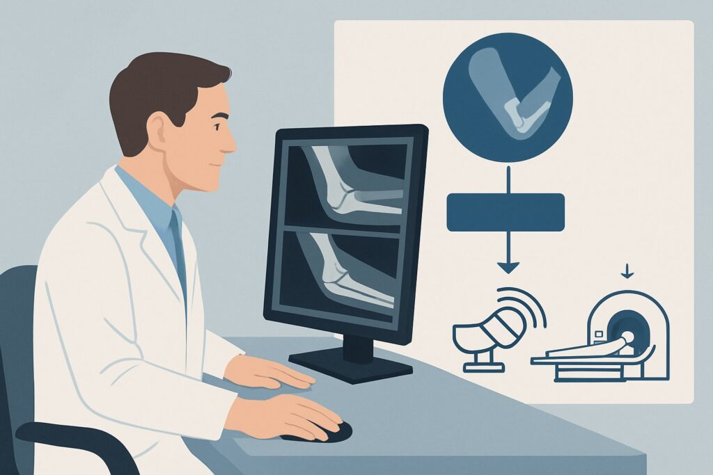
What’s the Next Step After Initial Elbow Radiographs?
The results of the initial elbow radiographs create a critical decision point in the clinical workflow, directing you toward either treatment or a more focused diagnostic plan.
If the radiograph is positive: When radiographs reveal a clear cause for the pain, such as moderate to severe osteoarthritis, a large loose body, or evidence of inflammatory arthritis, the diagnostic phase is often complete. The next step is to initiate clinical management. This may include activity modification, physical therapy, non-steroidal anti-inflammatory drugs (NSAIDs), corticosteroid injections, or referral to an orthopedic surgeon or rheumatologist for further management. Additional imaging is typically not needed at this stage.
If the radiograph is negative or shows non-specific findings: A normal radiograph is a valuable piece of information, as it effectively rules out significant bony pathology and narrows the differential diagnosis to soft-tissue or neurologic causes. At this point, you must return to the patient’s history and physical exam to refine the clinical question.
- If clinical suspicion for a tendon or ligament injury is high (e.g., focal tenderness over an epicondyle or collateral ligament), the next appropriate step is often an MRI or a focused musculoskeletal ultrasound. This aligns with the ACR variant for suspected chronic epicondylalgia or collateral ligament tear.
- If the patient’s symptoms are suggestive of nerve entrapment (e.g., cubital tunnel syndrome), the workup may proceed to MRI of the elbow to look for structural causes of compression, or to electrodiagnostic studies (EMG/NCS).
- If mechanical symptoms like locking are the primary complaint despite normal radiographs, an MR arthrogram may be considered to evaluate for non-ossified loose bodies or chondral defects.
In essence, a negative radiograph is not an endpoint but a gateway to a more targeted workup based on a refined clinical hypothesis.
Common Pitfalls to Avoid in the Initial Workup of Chronic Elbow Pain
Navigating the initial workup for chronic elbow pain requires avoiding several common missteps that can lead to delayed diagnosis or low-value care.
- Skipping Radiographs and Ordering MRI First: This is the most frequent pitfall. It often results in unnecessary expense and can reveal incidental soft-tissue findings that may not be the primary pain generator, while the true bony cause would have been easily identified on a simple radiograph.
- Over-attributing Symptoms to Mild OA: Finding mild degenerative changes on a radiograph does not automatically mean it is the cause of the patient’s symptoms. It is crucial to correlate the imaging findings with the clinical exam. Significant pain with minimal radiographic change should prompt consideration of a soft-tissue etiology.
- Stopping the Workup After a Negative Radiograph: A normal radiograph is a decision node, not a final diagnosis. Failing to proceed with a focused workup for soft-tissue or neurologic causes when clinically indicated can leave the patient without a clear diagnosis or treatment plan.
If red-flag symptoms are present—such as night pain, constitutional symptoms like fever or weight loss, or a palpable mass—escalate the workup promptly with advanced imaging (often MRI with contrast) and specialist consultation, as these may indicate a more serious underlying pathology like infection or malignancy.
Related ACR Topics and Tools
The ACR Appropriateness Criteria are a comprehensive resource for evidence-based imaging decisions. For breadth across all scenarios in Chronic Elbow Pain, see our parent guide: Chronic Elbow Pain: ACR Appropriateness Decoded.
For additional decision support and technical details, the following GigHz tools are available:
- ACR Appropriateness Criteria Lookup — To review guidelines for adjacent or alternative clinical scenarios.
- Imaging Protocol Library — For detailed technical specifications on performing various imaging studies.
- Radiation Dose Calculator — To help in discussions with patients about cumulative radiation exposure from medical imaging.
Frequently Asked Questions
Why not start with an MRI for chronic elbow pain, since it can see everything?
Starting with an MRI is considered ‘Usually not appropriate’ by the ACR for initial, undifferentiated chronic elbow pain. Radiographs are a much more cost-effective and efficient first step to rule out common bony causes like osteoarthritis. If radiographs are negative and a soft-tissue injury is still suspected, then an MRI becomes an appropriate next step.
If my patient has classic ‘tennis elbow’ symptoms, should I still order a radiograph first?
Yes, in most cases. While the diagnosis of lateral epicondylalgia is primarily clinical, a baseline radiograph is still recommended to rule out underlying bony pathology that could mimic or contribute to the symptoms. Advanced imaging like MRI or ultrasound is typically reserved for cases that are refractory to conservative treatment.
What specific views should I order for an initial elbow radiograph?
A standard elbow series typically includes anteroposterior (AP) and lateral views. These two projections provide a comprehensive initial assessment of the joint alignment, bone integrity, and articular surfaces. Oblique views may be added by the radiologist if there is a specific concern for a subtle fracture.
Is there any role for a CT scan in the initial workup of chronic elbow pain?
No, a CT scan is rated ‘Usually not appropriate’ for the initial evaluation. CT provides excellent detail of bone but involves more radiation than radiography and offers limited evaluation of soft tissues. Its role is typically reserved for problem-solving, such as better characterizing a complex fracture or a specific bony lesion identified on a prior radiograph.
What if the radiograph is negative but the patient’s pain is worsening?
A negative radiograph in the setting of worsening pain is a key indicator to proceed to the next step in the diagnostic workflow. Re-evaluate the patient’s symptoms and exam to form a more specific hypothesis (e.g., tendinopathy, ligament injury, nerve entrapment) and then order the appropriate advanced imaging study, most commonly an MRI.
Reviewed by Pouyan Golshani, MD, Interventional Radiologist — May 21, 2026