What Is the Right Initial Imaging for Chronic Rhinosinusitis Surgical Candidates?
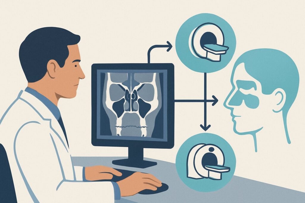
A 48-year-old patient is in your clinic after referral to Otolaryngology. He has endured four documented episodes of acute sinusitis in the past year, each treated with antibiotics, but his symptoms of facial pressure, nasal congestion, and post-nasal drip persist. He has failed maximal medical therapy and is now being considered for functional endoscopic sinus surgery (FESS). Before scheduling the procedure, the surgeon needs a detailed anatomical roadmap to plan the intervention. This article details the ACR-guided imaging workflow for a patient with acute recurrent or chronic rhinosinusitis being evaluated for surgery. For this specific scenario, the American College of Radiology notes that a CT maxillofacial without IV contrast is Usually Appropriate.
Who Fits This Clinical Scenario?
This guidance applies to patients with a diagnosis of chronic rhinosinusitis (CRS), defined as sinonasal inflammation lasting 12 weeks or longer, or acute recurrent rhinosinusitis (ARRS), defined as four or more episodes of acute bacterial rhinosinusitis per year without persistent symptoms between episodes. It also covers patients with related non-neoplastic conditions being considered for surgery, such as sinonasal polyposis, noninvasive fungal sinusitis, suspected mucocele, silent sinus syndrome, or a clinically significant deviated nasal septum.
The key differentiator for this workflow is the non-emergent, pre-surgical context. This guidance does not apply to patients with signs of complicated or invasive disease. Exclude patients from this pathway if they present with:
- Neurologic or orbital red flags: Proptosis, vision changes, ophthalmoplegia, severe headache, or altered mental status. These symptoms suggest potential orbital or intracranial complications and route to a different ACR workflow.
- Rapid progression or immunocompromise: A patient with uncontrolled diabetes or on immunosuppressive therapy who develops rapidly worsening symptoms is concerning for invasive fungal sinusitis, a medical emergency requiring a different imaging and management approach.
- Uncomplicated acute sinusitis: A patient with their first episode of sinusitis lasting less than four weeks, without complications, typically does not require any imaging.
What Diagnoses Are You Working Up in This Scenario?
The primary goal of imaging in this pre-surgical setting is to confirm the diagnosis, delineate the extent of disease, and map the patient’s unique sinonasal anatomy. The differential diagnosis includes several overlapping inflammatory conditions.
Chronic Rhinosinusitis (CRS) and Sinonasal Polyposis: This is the most common indication. Imaging aims to identify mucosal thickening, sinus opacification, and particularly, obstruction of the osteomeatal complex (OMC)—the final common pathway for drainage of the frontal, maxillary, and anterior ethmoid sinuses. In patients with polyposis, imaging will show smoothly marginated, polypoid soft tissue masses filling the nasal cavity and paranasal sinuses.
Noninvasive Fungal Sinusitis (Mycetoma): Also known as a fungus ball, this condition involves a tangled mass of fungal hyphae within a sinus cavity, most commonly the maxillary. On non-contrast CT, it can often be identified by the presence of hyperdense material (calcification or metallic-density material) within an opacified sinus.
Mucocele: This occurs when a sinus ostium is obstructed, leading to the accumulation of sterile mucus. Over time, the sinus expands, remodeling and thinning the bony walls. CT is excellent for showing this bony expansion and the homogeneously opacified sinus contents.
Silent Sinus Syndrome: A less common condition characterized by chronic maxillary sinus atelectasis. Obstruction of the infundibulum leads to negative pressure, causing the orbital floor to retract downward (enophthalmos) and the lateral nasal wall to bow medially. CT clearly demonstrates these characteristic bony changes.
Why Is CT Maxillofacial Without IV Contrast the Recommended Study?
For pre-surgical evaluation of chronic inflammatory sinonasal disease, a non-contrast CT of the maxillofacial region provides the ideal balance of diagnostic information and safety. The ACR rates this study as Usually Appropriate because it directly answers the key clinical questions.
The primary strength of CT is its superb spatial resolution and exquisite depiction of bony anatomy. For a surgeon planning FESS, this is non-negotiable. The scan provides a precise map of critical structures, including the uncinate process, lamina papyracea (the thin bone separating the ethmoid sinuses from the orbit), the skull base (cribriform plate and fovea ethmoidalis), and the location of the sphenoid sinus and its relationship to the optic nerves and carotid arteries. It also clearly defines the extent of mucosal thickening, air-fluid levels, and bony erosion or remodeling.
Intravenous contrast is not necessary in this scenario. The clinical question is about anatomy and inflammatory changes, not vascularity or enhancement patterns that would suggest a tumor or abscess—concerns that fall under different ACR scenarios. Omitting contrast avoids potential risks like allergic reaction and contrast-induced nephropathy while also reducing cost and scan time.
How do alternative studies compare?
- Radiography (plain films) are rated Usually not appropriate. They provide very limited detail due to overlapping structures, have poor sensitivity for mucosal disease, and offer no value for surgical planning.
- MRI orbits face neck without IV contrast is rated May be appropriate. While MRI offers superior soft-tissue contrast, it is significantly inferior to CT for evaluating the fine bony septa and landmarks essential for FESS. It is more costly, less accessible, and more susceptible to motion artifact. Its role is typically reserved for cases where CT findings are indeterminate or there is a specific concern for a soft tissue mass or intracranial extension.
The radiation dose for a maxillofacial CT is relatively low (adult relative radiation level ☢☢, 0.1-1 mSv), a key consideration in a patient population that may require follow-up imaging. While the detailed protocol varies by indication, our guide on non-contrast head CT covers related principles of technique and interpretation: CT Brain Without Contrast.
What’s Next After CT Maxillofacial Without Contrast? Downstream Workflow
The results of the non-contrast CT will directly guide the next steps in management, primarily confirming the need for and approach to surgery.
- If the study is positive for significant inflammatory disease: Findings of extensive mucosal thickening, osteomeatal complex obstruction, polyposis, or a mucocele confirm the diagnosis and provide the necessary anatomical detail for the surgeon to proceed with FESS. The specific surgical plan (e.g., maxillary antrostomy, total ethmoidectomy) will be tailored to the patient’s disease pattern and anatomy as shown on the CT.
- If the study is negative or shows only minimal disease: A normal CT scan in a symptomatic patient should prompt a re-evaluation of the diagnosis. The patient’s symptoms may stem from non-sinus causes like migraine, trigeminal neuralgia, or temporomandibular joint dysfunction. Further workup would be directed by these alternative considerations.
- If the study is indeterminate or suspicious for a mass: If the CT shows an unusual soft tissue pattern, unexpected bone destruction, or features not typical for inflammation, the workup pivots. An MRI, often with and without IV contrast, would be the next logical step to better characterize the soft tissues and assess for a potential sinonasal neoplasm. This moves the patient into the “Suspected sinonasal mass” ACR scenario.
Pitfalls to Avoid (and When to Get Help)
In this well-defined clinical scenario, several common pitfalls can lead to diagnostic errors or inappropriate management. First, avoid ordering imaging during an acute exacerbation of sinusitis unless a complication is suspected; active inflammation can overestimate the extent of chronic disease. It is best to obtain the scan after the patient has completed a course of medical therapy. Second, be careful not to misinterpret benign hyperdensity within a sinus as an invasive process; in the right context, it is a classic sign of a noninvasive mycetoma. Finally, ensure the scan protocol uses thin slices and appropriate reconstructions (coronal, sagittal, axial) to provide the surgeon with the necessary detail. If the CT report describes aggressive bone destruction or perineural spread, escalate immediately with a referral for contrast-enhanced MRI and biopsy.
Related ACR Topics and Tools
This article focuses on a single, common clinical scenario. For a comprehensive overview of imaging for all sinonasal conditions, from uncomplicated sinusitis to suspected CSF leaks, please refer to our parent guide. You can also use the tools below to explore other ACR criteria, review imaging protocols, and discuss radiation dose with your patients.
- For breadth across all scenarios in Sinonasal Disease, see our parent guide: Sinonasal Disease: ACR Appropriateness Decoded.
- ACR Appropriateness Criteria Lookup — for adjacent scenarios
- Imaging Protocol Library — for technique on the recommended study
- Radiation Dose Calculator — for cumulative dose conversations
Frequently Asked Questions
Why is a non-contrast CT preferred over a CT with contrast for pre-surgical chronic sinusitis?
A non-contrast CT is preferred because it provides excellent visualization of the bony anatomy and mucosal surfaces, which are the critical elements for surgical planning in chronic inflammatory disease. Intravenous contrast does not add significant information for these indications and introduces unnecessary risks (e.g., allergic reaction, nephrotoxicity) and cost. Contrast is reserved for when there is suspicion of a complication like an abscess or a tumor.
My patient has chronic sinusitis symptoms but the CT scan is normal. What should I do?
A normal sinonasal CT in a patient with facial pain or pressure should prompt consideration of alternative diagnoses. The differential includes migraine, cluster headaches, trigeminal neuralgia, temporomandibular joint (TMJ) dysfunction, and dental pathology. The clinical workup should be redirected to investigate these other potential causes.
When is an MRI a better choice than CT for sinonasal disease?
MRI is the preferred study when there is a clinical or CT-based suspicion of a sinonasal mass, perineural tumor spread, or intracranial/orbital complications. Its superior soft-tissue contrast is essential for characterizing tumors and their extent. For routine, uncomplicated chronic rhinosinusitis evaluation before surgery, CT remains the primary modality due to its superior bony detail.
Should I order imaging for a patient with their first episode of acute sinusitis?
No. For uncomplicated acute rhinosinusitis (symptoms <4 weeks without red flags), imaging is rated as 'Usually Not Appropriate' by the ACR. The diagnosis is clinical, and imaging does not change management. Imaging is only indicated in the acute setting if you suspect a complication, such as orbital cellulitis or an intracranial abscess.
What is the role of Cone Beam CT (CBCT) in this scenario?
Cone Beam CT is rated as ‘May be appropriate’ by the ACR for this indication. It is an alternative to conventional CT that can provide high-resolution images of the sinonasal anatomy, often with a lower radiation dose. It is frequently used in otolaryngology and dental offices. However, its soft-tissue contrast resolution is generally lower than that of multidetector CT (MDCT), which may be a limitation in some cases.
Reviewed by Pouyan Golshani, MD, Interventional Radiologist — May 29, 2026