What Is the Right Initial Imaging for Nonmassive Hemoptysis? An ACR-Guided Workflow
A 52-year-old man with a 30-pack-year smoking history presents to your clinic after noticing streaks of blood in his sputum for the past two days. He is hemodynamically stable, with no shortness of breath or chest pain. He estimates the total volume is less than a teaspoon. You need to begin a diagnostic workup, but which imaging study is the most appropriate first step to evaluate this non–life-threatening hemoptysis? This article provides a focused clinical workflow for this specific scenario, explaining why the American College of Radiology (ACR) designates a chest radiograph as Usually Appropriate for the initial evaluation.
Who Fits This Clinical Scenario for Nonmassive Hemoptysis?
This guidance applies to patients presenting with nonmassive (non–life-threatening) hemoptysis for their initial imaging workup. The key inclusion criteria are hemodynamic stability and a small volume of bleeding. While definitions vary, nonmassive hemoptysis is generally considered to be less than 100 mL of expectorated blood over a 24-hour period. The patient should not be in any respiratory distress, and their vital signs should be normal.
It is critical to distinguish this presentation from similar but distinct clinical situations that require a different imaging approach:
- Exclusion 1: Massive Hemoptysis. If the patient is hemodynamically unstable, has significant respiratory compromise, or is coughing up large volumes of blood (often defined as >100-600 mL/24 hours), this constitutes a medical emergency. This presentation falls under the Massive (life-threatening) hemoptysis scenario, which typically requires immediate multidetector CT angiography (CTA) and consultation with interventional radiology and/or thoracic surgery.
- Exclusion 2: Recurrent Hemoptysis. If the patient has had multiple, distinct episodes of hemoptysis, especially after a prior negative or inconclusive workup, the imaging strategy changes. This falls into the Recurrent hemoptysis scenario, where CT is often the preferred initial modality, as a chest radiograph may have insufficient sensitivity for subtle underlying causes.
This article’s workflow is designed for the common, stable, first-time presentation of scant hemoptysis seen in an outpatient or emergency department setting.
What Diagnoses Are You Working Up in Nonmassive Hemoptysis?
The initial imaging choice is guided by a differential diagnosis that spans from benign, self-limiting conditions to life-threatening diseases. The goal is to efficiently identify or exclude the most common and most serious etiologies.
Airway Inflammation and Infection
By far the most common cause of nonmassive hemoptysis is inflammation of the airways. Acute or chronic bronchitis can cause mucosal irritation and friability, leading to blood-streaked sputum. Pneumonia is another frequent cause, where inflammation of the lung parenchyma can result in bleeding. Imaging in this context seeks to identify a focal infiltrate, consolidation, or signs of bronchial wall thickening that would support an infectious or inflammatory diagnosis.
Bronchogenic Carcinoma
In patients with risk factors, particularly a history of smoking, lung cancer is a critical diagnosis to exclude. A tumor eroding into a bronchial vessel can present with hemoptysis, which may be the first and only symptom. The initial imaging study is a crucial screening tool for a pulmonary mass, nodule, or hilar/mediastinal adenopathy that would prompt further investigation with advanced imaging and biopsy.
Bronchiectasis
This condition involves permanent, abnormal dilation of the bronchi, often resulting from recurrent infections or chronic inflammation. The dilated airways have hypertrophied and fragile bronchial arteries running alongside them, which are prone to rupture and bleeding. Imaging aims to detect characteristic findings like bronchial wall thickening, lack of normal bronchial tapering (“tram tracking”), and cystic changes.
Other Less Common Causes
While less frequent, other important considerations include diffuse alveolar hemorrhage (often from autoimmune conditions like vasculitis), pulmonary embolism, arteriovenous malformations (AVMs), and tuberculosis. The initial imaging study may reveal findings suggestive of these diagnoses, such as diffuse ground-glass opacities in DAH or a wedge-shaped opacity in pulmonary infarction from a PE.
Why Is a Chest Radiograph the Recommended First Step for This Presentation?
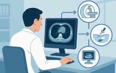
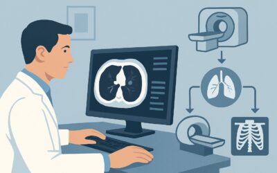
For the initial evaluation of nonmassive hemoptysis, the ACR Appropriateness Criteria panel rates Radiography chest as Usually Appropriate. This recommendation is based on a careful balance of diagnostic yield, accessibility, cost, and patient safety.
A standard two-view (posteroanterior and lateral) chest radiograph serves as an excellent first-line screening tool. It is highly effective at identifying many of the most common and serious causes in the differential, including pneumonia, a large lung mass, significant atelectasis, or extensive bronchiectasis. Its wide availability and low cost make it an efficient starting point for the workup. A positive finding can immediately guide subsequent management, while a negative result in a low-risk patient can provide reassurance and justify a course of observation.
The radiation dose is a key advantage. A chest radiograph delivers a very low effective dose (adult RRL ☢ <0.1 mSv), minimizing the patient’s exposure to ionizing radiation. This contrasts sharply with the alternatives.
Comparison to Other Modalities:
- CT chest with IV contrast is also rated Usually Appropriate but is not typically the first-line study. It offers far greater detail and sensitivity for subtle nodules, bronchiectasis, and vascular abnormalities. However, it comes with a substantially higher radiation dose (adult RRL ☢☢☢ 1-10 mSv) and the risks associated with intravenous contrast. It is best reserved as a second-line study if the chest radiograph is negative in a high-risk patient or if a specific finding on the radiograph requires further characterization.
- CT chest without and with IV contrast is rated Usually not appropriate. Performing both a non-contrast and a contrast-enhanced scan for this initial indication needlessly doubles the radiation dose compared to a single contrast-enhanced acquisition. The pre-contrast images rarely add diagnostically crucial information that would change management in this specific scenario.
The logical starting point is the test that answers the most clinical questions with the least risk and cost. For nonmassive hemoptysis, that test is the chest radiograph.
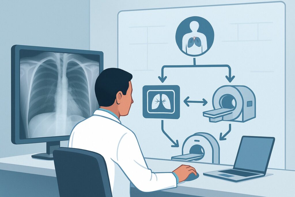
What’s Next After a Chest Radiograph? Downstream Workflow
The results of the initial chest radiograph will dictate the subsequent diagnostic and management pathway. The workflow branches based on whether the study is positive, negative, or equivocal, and must be interpreted in the context of the patient’s risk factors (e.g., age, smoking history).
If the Chest Radiograph is Positive:
- Finding suggests infection (e.g., focal infiltrate): The appropriate next step is to initiate antibiotic therapy and arrange for follow-up imaging after treatment to ensure resolution.
- Finding suggests malignancy (e.g., mass, nodule, adenopathy): The patient should be referred for a CT of the chest with IV contrast to better characterize the lesion, evaluate for metastatic disease, and guide a potential biopsy.
- Finding suggests bronchiectasis or other chronic lung disease: Management should focus on the underlying condition. A non-contrast high-resolution CT (HRCT) may be considered for better anatomic detail of bronchiectasis if needed.
If the Chest Radiograph is Negative:
- In a low-risk patient (e.g., under 40, non-smoker, clear infectious trigger): A period of clinical observation is often appropriate. If the hemoptysis resolves, no further workup may be necessary.
- In a high-risk patient (e.g., age >40 with a significant smoking history) or if hemoptysis persists or recurs: A negative radiograph is not sufficient to rule out pathology. The next step is to proceed with a CT chest with IV contrast. CT is significantly more sensitive for detecting small endobronchial lesions, subtle bronchiectasis, and arteriovenous malformations that are commonly missed on plain films. If symptoms recur after a negative CT, the patient may fit the “Recurrent hemoptysis” scenario, potentially requiring bronchoscopy.
Pitfalls to Avoid (and When to Get Help)
Navigating the workup for nonmassive hemoptysis requires careful clinical judgment. Here are several common pitfalls to avoid:
- Stopping the workup too early: Do not dismiss hemoptysis in a high-risk patient (smoker >40) just because the initial chest radiograph is normal. A significant percentage of patients with lung cancer have a normal chest X-ray at presentation.
- Misclassifying the severity: Underestimating the volume of bleeding can lead to delaying a more aggressive workup. If there is any doubt about hemodynamic stability or the rate of bleeding, manage the patient as if they have massive hemoptysis.
- Forgetting the upper airway and GI tract: Ensure the bleeding is truly from the lower respiratory tract (hemoptysis) and not from the nasopharynx (epistaxis) or the gastrointestinal tract (hematemesis).
- Ignoring risk factors: The pre-test probability of serious disease heavily influences the downstream workflow. The same negative chest radiograph has very different implications for a 25-year-old with a viral URI versus a 65-year-old smoker.
If the patient’s condition worsens, if bleeding becomes massive, or if the diagnosis remains elusive after non-invasive imaging, escalate care by consulting with Pulmonology for consideration of bronchoscopy and/or Interventional Radiology for potential CTA and embolization.
Related ACR Topics and Tools
The ACR Appropriateness Criteria are a powerful resource for evidence-based imaging decisions. For a comprehensive overview of all clinical variants related to hemoptysis, from massive to recurrent, please see our parent topic hub article. For tools to help with ordering, protocoling, and discussing radiation dose, see the resources below.
- For breadth across all scenarios in Hemoptysis, see our parent guide: Hemoptysis: ACR Appropriateness Decoded.
- To look up other clinical scenarios, visit the ACR Appropriateness Criteria Lookup tool.
- For detailed technical parameters of imaging studies, explore the Imaging Protocol Library.
- To help explain radiation exposure to patients, use the Radiation Dose Calculator.
Frequently Asked Questions
Is a CT scan always necessary if the initial chest X-ray is negative for hemoptysis?
Not always. For a low-risk patient (e.g., young, non-smoker, with a likely infectious cause like bronchitis), if the hemoptysis is minor and resolves, clinical observation after a negative chest X-ray may be sufficient. However, for a high-risk patient (e.g., over 40 with a significant smoking history) or if symptoms persist, a CT chest with IV contrast is the recommended next step because of its higher sensitivity for detecting underlying causes like small tumors or bronchiectasis.
Why is CT with and without contrast ‘Usually Not Appropriate’ for this initial workup?
A combined ‘with and without contrast’ CT study, also known as a multiphase scan, delivers a double dose of radiation compared to a single contrast-enhanced scan. For the initial evaluation of nonmassive hemoptysis, the pre-contrast (non-contrast) images rarely provide additional diagnostic information that would change management. A single, well-timed contrast-enhanced CT is sufficient to evaluate the lung parenchyma, airways, and mediastinal and hilar structures.
What if the patient has renal insufficiency and cannot receive IV contrast?
If a CT is indicated (e.g., high-risk patient with a negative chest radiograph) but the patient has a contraindication to IV contrast, a CT chest without IV contrast is rated as ‘May be appropriate’ by the ACR. While less sensitive for vascular abnormalities and some tumors, a non-contrast high-resolution CT can still provide excellent detail of the lung parenchyma and airways, and is very effective for diagnosing bronchiectasis, emphysema, or interstitial lung disease.
How does this workflow change if the hemoptysis is massive and life-threatening?
The workflow changes dramatically. Massive hemoptysis is a medical emergency requiring immediate stabilization of the airway, breathing, and circulation (ABCs). The ACR recommends CTA chest with IV contrast as the initial imaging study, often bypassing the chest radiograph entirely. The goal of CTA is to rapidly identify the source of bleeding (typically a hypertrophied bronchial artery) to guide immediate intervention, such as bronchial artery embolization by interventional radiology.
Should I order a lateral view with the PA chest radiograph?
Yes. A two-view chest radiograph (posteroanterior and lateral) is the standard of care. The lateral view is essential for evaluating regions that are obscured on the PA view, such as the retrosternal space, the retrocardiac region, and the posterior costophrenic sulci. Omitting the lateral view can lead to missed pathology, including lung nodules or infiltrates.
Reviewed by Pouyan Golshani, MD, Interventional Radiologist — May 29, 2026