What Is the Right Initial Imaging for Total Knee Arthroplasty Follow-Up?
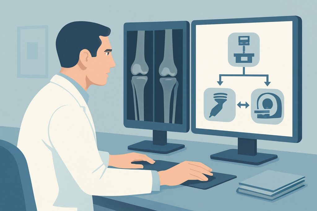
A 72-year-old patient, five years out from a total knee arthroplasty (TKA), presents to your clinic for a routine follow-up. He mentions some vague, intermittent anterior knee pain but no acute symptoms. Another patient, 6 years post-TKA, is entirely asymptomatic but is due for surveillance. In both cases, the clinical question is the same: what is the appropriate initial imaging study to assess the status of the prosthesis and the surrounding bone? This decision point is common in primary care and orthopedic clinics, balancing the need for surveillance against unnecessary radiation and cost. For this specific scenario—the initial imaging for either symptomatic or asymptomatic TKA follow-up—the American College of Radiology (ACR) designates Radiography knee as Usually appropriate. This article provides a detailed workflow for this common clinical crossroads.
Who Fits This Clinical Scenario for Initial TKA Follow-up?
This guidance applies to any patient with a total knee arthroplasty who requires initial imaging, whether for routine surveillance in an asymptomatic patient or for the evaluation of new or chronic, non-acute symptoms like pain, stiffness, or a feeling of instability. The key qualifier is that this is the initial imaging step for the current clinical presentation. The patient has not had other recent, relevant imaging for this specific complaint.
This workflow is distinct from several related but more urgent clinical situations. It does not apply to patients with:
- Strong suspicion of acute infection: Patients presenting with fever, chills, significant localized erythema, warmth, or purulent drainage from the surgical site require a different, more urgent workup, often involving laboratory tests (ESR, CRP) and potentially joint aspiration before or alongside imaging. While radiographs are still obtained, the subsequent imaging pathway is different.
- High-energy trauma or acute fracture suspicion: A patient with a TKA who suffers a significant fall or direct trauma, where an acute periprosthetic fracture is the primary concern, may follow a modified imaging protocol.
- A specific, isolated clinical question: If the workup has already progressed and the goal is to answer a focused question—such as measuring component rotation or evaluating a suspected soft-tissue abnormality after initial radiographs are complete—those scenarios are covered by different ACR Appropriateness Criteria variants.
This article focuses squarely on the first imaging test to order when evaluating the status of a TKA in a non-emergent setting.
What Diagnoses Are You Working Up with Initial TKA Imaging?
The initial radiograph in a TKA follow-up serves as a crucial screening and surveillance tool, aiming to detect the most common modes of arthroplasty failure and other complications. The differential diagnosis is broad and includes both mechanical and biological issues.
The most common long-term complication you are assessing for is aseptic loosening. This occurs when the bond between the implant and the bone weakens over time, without an infectious cause. Radiographs are the primary modality for detecting the hallmark sign of loosening: progressive radiolucent lines at the bone-cement or bone-implant interface.
Another key diagnosis is osteolysis, or “particle disease.” This is a biological process where microscopic wear debris from the polyethylene liner incites an inflammatory response, leading to bone resorption around the implant. On radiographs, this appears as focal, well-defined areas of bone loss (lytic lesions), which can compromise implant stability and lead to fracture.
You are also evaluating for component wear and positioning. While the polyethylene liner itself is radiolucent, indirect signs of wear can be seen, such as asymmetric joint space narrowing or changes in component alignment. Gross malposition, subsidence (sinking) of a component, or significant changes in overall limb alignment are also readily assessed on weight-bearing films.
Less commonly, a subtle, non-displaced periprosthetic fracture may be the cause of new pain. Radiographs are the first-line study to identify fracture lines, though they can sometimes be difficult to see, especially around the metallic components. Finally, radiographs establish a valuable baseline for future comparison, which is critical for determining the stability or progression of any subtle findings over time.
Why Are Knee Radiographs the Recommended Initial Study for TKA Follow-up?
The ACR rates Radiography knee as Usually appropriate for this scenario because it is a high-yield, low-risk, and cost-effective examination that directly addresses the most common differential diagnoses. Standard knee radiographs provide excellent visualization of the bone, the metallic components, and the critical interface between them.
Weight-bearing anteroposterior (AP), lateral, and skyline (patellar) views are the standard of care. These views are essential for assessing component alignment, stability under load, joint space (as a surrogate for polyethylene wear), and the presence of radiolucent lines or osteolysis that could indicate loosening. The low relative radiation level (ACR RRL ☢ <0.1 mSv) makes it a safe tool for serial follow-up over the lifetime of the implant. Its wide availability and low cost make it the most practical and efficient first step. In contrast, more advanced imaging modalities are rated Usually not appropriate as the initial study:
- MRI knee (without or with IV contrast): While specialized metal artifact reduction sequences (MARS) have improved MRI’s utility in evaluating prosthetic joints, it is not a first-line tool. It is more expensive, less accessible, and often degraded by artifact, making it difficult to assess the bone-implant interface—the primary goal of initial follow-up. MRI is reserved for specific downstream questions, such as evaluating for soft-tissue abnormalities like synovitis or muscle tears.
- CT knee: CT provides excellent cross-sectional detail of the bone and can be useful for assessing osteolysis, component position, and occult fractures. However, it involves more radiation than radiography (ACR RRL ☢ <0.1 mSv) and is also susceptible to metal artifact. Its primary role is as a second-line test for specific indications, such as measuring component rotation when malalignment is suspected after initial radiographs.
For the initial evaluation of a TKA, radiographs provide the necessary information to guide management in the vast majority of cases, making it the clear and appropriate starting point.
What’s Next After Knee Radiographs? Downstream Workflow
The results of the initial knee radiographs dictate the subsequent clinical pathway. The decision tree branches based on the findings in the context of the patient’s symptoms.
If radiographs are normal and the patient is asymptomatic: This is the ideal outcome for a routine surveillance exam. No further imaging is needed. The patient can continue with routine clinical follow-up as determined by their orthopedic surgeon, typically every few years.
If radiographs are normal but the patient remains symptomatic: This is a common and challenging scenario. The absence of radiographic findings does not rule out a problem. The next step is a deeper clinical and laboratory evaluation to rule out low-grade infection, with tests like Erythrocyte Sedimentation Rate (ESR) and C-Reactive Protein (CRP). If these are elevated, joint aspiration may be warranted. If infection is excluded, the workup may proceed to advanced imaging to investigate other causes of pain, such as component malrotation (CT scan) or soft-tissue pathology (MARS MRI). This pathway aligns with other ACR variants, such as “Pain after total knee arthroplasty. Infection excluded.”
If radiographs show clear evidence of loosening, osteolysis, or component failure: This patient should be referred promptly to an orthopedic surgeon. These findings often represent mechanical failure of the arthroplasty and may require revision surgery. The surgeon may order additional imaging, such as a CT scan, for pre-operative planning, but the initial radiograph has successfully identified the problem and guided the primary management decision.
If radiographs are indeterminate or show subtle, non-specific changes: The most critical action is to obtain and compare with all available prior radiographs. A subtle radiolucent line that is new or progressing is far more significant than one that has been stable for years. If no priors are available or the findings remain equivocal, a follow-up radiograph in 3-6 months may be appropriate to assess for change.
Common Pitfalls in Initial TKA Imaging (and When to Escalate)
To maximize the diagnostic value of initial TKA imaging, it is important to avoid several common pitfalls.
First, failing to order weight-bearing views is a significant error. Non-weight-bearing films can mask subtle instability, malalignment, and asymmetric polyethylene wear. Always specify “weight-bearing AP” view when ordering.
Second, evaluating images in a vacuum without prior films can lead to misinterpretation. What might appear to be a concerning radiolucent line could be a stable, non-progressive finding. Always make an effort to retrieve and compare with previous studies.
Third, over-relying on radiographs to rule out infection is a critical mistake. Radiographs are highly insensitive for early periprosthetic joint infection (PJI). A normal radiograph does not exclude PJI. If there is any clinical suspicion of infection (persistent pain, effusion, low-grade fever), the workup must include inflammatory markers (ESR/CRP).
If a patient presents with acute, severe pain, constitutional symptoms, or obvious signs of infection, escalate immediately with laboratory workup and an urgent orthopedic consultation. Do not delay this for a routine imaging appointment.
Related ACR Topics and Tools
For a comprehensive overview of all clinical scenarios related to imaging after a total knee arthroplasty, please refer to our parent guide. Additional GigHz resources can help refine your imaging decisions and technique.
- For breadth across all scenarios in Imaging After Total Knee Arthroplasty, see our parent guide: Imaging After Total Knee Arthroplasty: ACR Appropriateness Decoded.
- To explore other clinical presentations, use the ACR Appropriateness Criteria Lookup.
- For details on specific imaging techniques, consult the Imaging Protocol Library.
- To discuss radiation exposure with patients, the Radiation Dose Calculator can be a useful tool.
Frequently Asked Questions
How often should an asymptomatic patient with a total knee arthroplasty get follow-up radiographs?
There is no universal consensus, but many orthopedic societies recommend surveillance radiographs at intervals, such as at 1, 3, 5, and then every 2-5 years thereafter. The goal is to detect potential issues like osteolysis or wear before they become symptomatic or lead to catastrophic failure. The exact schedule should be determined by the patient’s surgeon.
What specific radiographic views are most important for a TKA follow-up?
The standard series includes a weight-bearing anteroposterior (AP) view, a lateral view, and a skyline (or sunrise/merchant) view of the patella. The weight-bearing AP is critical for assessing alignment and joint space, the lateral view assesses flexion/extension and anteroposterior component position, and the skyline view is essential for evaluating the patellofemoral articulation.
If radiographs are negative but my patient has persistent knee pain, what is the next step?
A negative radiograph in a symptomatic patient warrants further investigation. The first step is to rule out infection with laboratory tests (ESR and CRP). If those are negative, the differential includes soft-tissue impingement, instability not visible on static films, or component malrotation. Depending on the specific symptoms, the next imaging study might be a CT scan to assess component rotation or a MARS protocol MRI to evaluate soft tissues.
Can radiographs directly show polyethylene liner wear?
No, the polyethylene liner is radiolucent, meaning it is not visible on X-rays. However, radiographs can show indirect signs of wear. As the polyethylene thins, the space between the femoral and tibial components will narrow. Asymmetric narrowing on a weight-bearing film is a strong indicator of liner wear.
Is a bone scan a good initial test for a painful TKA?
No, a 3-phase bone scan is rated ‘Usually not appropriate’ by the ACR for the initial imaging of a TKA. While it is very sensitive for detecting increased metabolic activity (which can be seen in loosening or infection), it is not specific. Increased uptake around a prosthesis can persist for up to 1-2 years after surgery normally. It is considered a second or third-line test for problem-solving in complex cases, not for initial evaluation.
Reviewed by Pouyan Golshani, MD, Interventional Radiologist — May 30, 2026