Which Imaging Best Characterizes a Liver Lesion in a Patient with Known Malignancy?
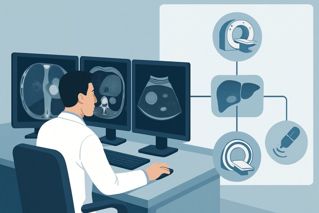
An oncologist reviews a surveillance scan for a 62-year-old patient with a history of resected colon cancer. The report for the single-phase portal venous Computed Tomography (CT) of the abdomen and pelvis is clean, except for one finding: a new, 1.8 cm low-density lesion in the right hepatic lobe. It doesn’t have the clear characteristics of a simple cyst, and on this limited study, its nature is indeterminate. The immediate clinical question is whether this represents metastatic disease, which would significantly alter the patient’s prognosis and management. This article provides a step-by-step clinical workflow for this exact scenario, guiding the choice of the next imaging study. According to the American College of Radiology (ACR) Appropriateness Criteria, an `MRI abdomen without and with IV contrast` is rated Usually appropriate as the next diagnostic step.
Who Fits This Clinical Scenario for an Indeterminate Liver Lesion?
This guidance is specifically for patients who meet a precise set of criteria. Applying this workflow to the wrong clinical context can lead to diagnostic delays or unnecessary testing. This article applies to a patient who has:
- A known history of an extrahepatic malignancy (e.g., colorectal, breast, lung, or pancreatic cancer). The pre-test probability of metastasis is a critical factor.
- An indeterminate liver lesion greater than 1 cm in size.
- The lesion was discovered on an initial imaging study that was insufficient for full characterization. This includes a noncontrast CT, a single-phase contrast-enhanced CT (e.g., portal venous phase only), or a noncontrast Magnetic Resonance Imaging (MRI).
Conversely, this workflow does not apply if the clinical situation is different. For example:
- No history of malignancy: If the patient has no known cancer, the differential diagnosis and imaging approach change, as the concern for metastasis is much lower.
- Known chronic liver disease or cirrhosis: In these patients, the primary concern shifts to hepatocellular carcinoma (HCC), and the workup follows a different pathway, often using the LI-RADS (Liver Imaging Reporting and Data System).
- Lesion is less than 1 cm: Small lesions are often followed with surveillance imaging rather than an immediate, aggressive workup, as they are frequently benign and difficult to characterize definitively.
- Initial study was a multiphase liver CT or MRI: If a high-quality, dedicated liver imaging study has already been performed, the lesion is not “indeterminate” in the same way; the next step is based on that definitive report, not re-characterization.
What Diagnoses Are You Working Up in This Scenario?
In a patient with a known extrahepatic cancer, any new, indeterminate liver lesion must be considered a metastasis until proven otherwise. The entire diagnostic algorithm is built around confirming or refuting this primary concern. However, benign lesions are common in the general population, and their presence can mimic metastatic disease, making a robust differential diagnosis essential.
Metastasis
This is the most consequential diagnosis and the primary driver for further imaging. The liver’s dual blood supply and filtration role make it a common site for hematogenous spread from many primary tumors, particularly those of the gastrointestinal tract (like colon cancer), lung, and breast. The appearance of metastases can be variable—hypovascular, hypervascular, cystic, or calcified—which is why a limited initial scan often leaves them indeterminate.
Benign Lesions (e.g., Hemangioma, FNH, Adenoma)
Even in patients with cancer, incidental benign liver lesions are frequently encountered. A cavernous hemangioma is the most common benign liver tumor, characterized by specific enhancement patterns (peripheral, nodular, centripetal fill-in) that are best seen on multiphase imaging. Focal Nodular Hyperplasia (FNH) is another common benign lesion, often containing a central scar. Differentiating these from hypervascular metastases (from primaries like renal cell carcinoma or neuroendocrine tumors) is a key goal of the workup.
Primary Liver Malignancy (e.g., HCC)
While less common in a patient without underlying chronic liver disease, a primary liver cancer like hepatocellular carcinoma can occur. It is a more significant consideration if the patient has risk factors like viral hepatitis or alcohol use, even if cirrhosis is not yet established. Distinguishing HCC from metastasis is critical as treatments differ substantially.
Abscess or Other Inflammatory Lesion
Though less likely to be the top consideration without corresponding clinical signs like fever or leukocytosis, a developing hepatic abscess can appear as an indeterminate complex cystic or enhancing lesion. The patient’s clinical status and recent history are vital clues in considering this diagnosis.
Why Is MRI Abdomen Without and With IV Contrast the Recommended Study?
For this specific clinical scenario, the ACR rates `MRI abdomen without and with IV contrast` as Usually appropriate. This recommendation is based on MRI’s superior soft tissue contrast and its ability to characterize lesions using multiple imaging sequences and contrast dynamics without exposing the patient to ionizing radiation.
The strength of a multiphase liver MRI lies in its comprehensive approach. Pre-contrast sequences (T1- and T2-weighted images) provide initial characterization; for example, a simple cyst will be very bright on T2 images, while a hemangioma is classically “lightbulb” bright. The dynamic, post-contrast portion of the exam, using a gadolinium-based contrast agent, is crucial. Images are acquired in late arterial, portal venous, and delayed phases. This allows the radiologist to assess the lesion’s vascularity and contrast washout patterns, which are key to differentiating metastases from benign entities like FNH or hemangiomas. The addition of hepatobiliary-specific contrast agents can further increase diagnostic confidence by showing whether a lesion contains functioning hepatocytes (as seen in FNH) or not (as in metastases or adenomas).
Why are other studies rated lower for this specific task?
- A `CT abdomen with IV contrast multiphase` is also rated Usually appropriate and is a valid alternative, especially if MRI is unavailable or contraindicated. However, it delivers a significant radiation dose (☢☢☢☢ 10-30 mSv) and has slightly lower sensitivity for detecting and characterizing small lesions compared to MRI.
- An `Image-guided biopsy liver` is rated May be appropriate. While it provides a definitive tissue diagnosis, it is an invasive procedure with risks of bleeding, infection, and tumor seeding. Biopsy is typically reserved for cases where high-quality imaging remains indeterminate or when a tissue diagnosis is required to guide systemic therapy. The goal of noninvasive imaging is to avoid biopsy whenever possible.
When ordering the recommended study, it is crucial to provide the radiologist with the patient’s specific history of malignancy. This context is essential for protocoling the MRI correctly and interpreting the findings accurately. Specifying “liver lesion characterization” on the order helps ensure the appropriate multiphase contrast sequences are performed.
What’s Next After MRI Abdomen Without and With IV Contrast? Downstream Workflow
The results of the liver MRI will guide the subsequent management plan, which typically branches into one of three pathways. The overarching goal is to clarify the patient’s disease stage and determine the appropriate next therapeutic step.
If the MRI confirms metastasis:
A definitive finding of one or more liver lesions characteristic of metastases confirms Stage IV disease. The next step is typically consultation with a multidisciplinary tumor board. Further systemic imaging, such as an `FDG-PET/CT skull base to mid-thigh` (rated Usually appropriate in this scenario), may be performed to assess the full extent of metastatic disease throughout the body. The patient’s treatment plan will likely be adjusted to include systemic chemotherapy, targeted therapy, or immunotherapy. In select cases of limited (oligometastatic) disease, local treatments like surgical resection, thermal ablation, or stereotactic body radiation therapy (SBRT) may be considered.
If the MRI confirms a benign lesion:
If the MRI confidently identifies the lesion as a classic hemangioma, FNH, or simple cyst, the workup for that finding is complete. This is a significant and reassuring result, as it means the patient’s cancer has not spread to the liver. No further imaging or follow-up for the benign lesion is typically required. The patient can continue with their established cancer surveillance and treatment plan without modification due to this finding.
If the MRI remains indeterminate:
In a small number of cases, even a high-quality MRI may not yield a definitive diagnosis. The lesion may have atypical features that overlap between benign and malignant entities. In this situation, the next step is often a discussion between the oncologist and radiologist. Options include short-term follow-up imaging (e.g., a repeat MRI in 3 months) to assess for stability or growth, proceeding to `Image-guided biopsy liver` (May be appropriate) for a tissue diagnosis, or utilizing a problem-solving modality like FDG-PET/CT, which can help differentiate metabolically active metastases from inactive benign lesions.
Pitfalls to Avoid (and When to Get Help)
Navigating this diagnostic pathway requires careful attention to detail to avoid common errors that can delay diagnosis or lead to unnecessary procedures.
- Pitfall 1: Repeating the wrong study. Ordering another single-phase CT after the first one was indeterminate will not solve the problem. The key is to escalate to a more definitive, multiphase characterization study like MRI or a multiphase CT.
- Pitfall 2: Omitting the clinical history. Failing to state “known history of [specific] cancer” on the imaging requisition is a critical error. This information allows the radiologist to tailor the imaging protocol and apply the correct interpretive lens.
- Pitfall 3: Prematurely ordering a biopsy. Jumping directly to a biopsy before obtaining high-quality, noninvasive characterization imaging exposes the patient to unnecessary risks. A definitive diagnosis can often be made with MRI alone.
- Pitfall 4: Misinterpreting stability. In a patient on active systemic therapy, a metastatic lesion may not grow. Stability on a short-term follow-up scan does not definitively rule out malignancy in this context.
If the results of a high-quality MRI are equivocal or conflict with the overall clinical picture, it is time to escalate. A discussion with the interpreting radiologist or presentation at a multidisciplinary tumor board is the appropriate next step to form a consensus plan.
Related ACR Topics and Tools
This article focuses on one specific clinical scenario. For a comprehensive overview of all variants related to the initial workup of a liver lesion, or to explore the tools used in this process, the following resources are available.
- For breadth across all scenarios in Liver Lesion-Initial Characterization, see our parent guide: Liver Lesion-Initial Characterization: ACR Appropriateness Decoded.
- ACR Appropriateness Criteria Lookup — To review the evidence for other, related clinical presentations.
- Imaging Protocol Library — For detailed technical specifications on how studies like a multiphase liver MRI are performed.
- Radiation Dose Calculator — To help in discussions with patients about cumulative radiation exposure from different imaging strategies.
Frequently Asked Questions
Why not just go straight to a PET/CT for a patient with known cancer?
While FDG-PET/CT is rated *Usually appropriate* and is excellent for systemic staging, it is not the primary tool for initial lesion characterization. MRI provides superior anatomical detail and tissue-specific information (like washout patterns) that are often more definitive for diagnosing the specific type of liver lesion. PET/CT is often used as the next step after a lesion is confirmed to be metastatic on MRI or CT to look for disease elsewhere.
What if my patient has a contraindication to MRI, like a non-compatible pacemaker?
If MRI is contraindicated, the ACR-recommended alternative is a `CT abdomen with IV contrast multiphase`, which is also rated *Usually appropriate*. This study uses a timed, multi-scan approach after IV contrast injection to assess the lesion’s enhancement pattern across arterial, portal venous, and delayed phases, providing a robust characterization, albeit with ionizing radiation and slightly lower soft tissue resolution than MRI.
Does the type of primary cancer matter when choosing the imaging study?
Yes, the primary cancer type provides crucial context. For instance, with a known neuroendocrine tumor, a `DOTATATE PET/CT` (*May be appropriate*) might be considered as it specifically targets somatostatin receptors. For most common epithelial cancers (colon, lung, breast), a standard multiphase MRI or CT is the standard characterization tool. Always include the primary cancer type in the imaging order.
What if the lesion was found on ultrasound instead of CT or MRI?
That is a different clinical scenario. If an indeterminate lesion >1 cm is found on ultrasound in a patient with known malignancy, the next step is often still a definitive cross-sectional study. Both multiphase CT and multiphase MRI are considered *Usually appropriate* in that specific context, with the choice depending on local expertise and patient factors.
Is contrast-enhanced ultrasound (CEUS) an option?
Contrast-enhanced ultrasound (`US abdomen with IV contrast`) is rated *May be appropriate* by the ACR for this scenario. It is a valuable, radiation-free tool for lesion characterization, particularly in patients with contraindications to both CT and MRI contrast agents. However, its availability and the local expertise required for performance and interpretation are more limited than for CT and MRI, making it a secondary option in many centers.
Reviewed by Pouyan Golshani, MD, Interventional Radiologist — May 29, 2026