Which Imaging Is Best for High-Risk Breast Cancer Screening in Women Under 30?

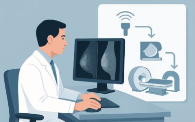
A 28-year-old woman with a known BRCA1 pathogenic variant presents to establish care and discuss her breast cancer screening plan. She is asymptomatic and has no personal history of breast cancer, but her mother and aunt were both diagnosed in their early 40s. You know that her high-risk status necessitates starting surveillance well before the standard age of 40, but the optimal imaging strategy in such a young patient, with predictably dense breast tissue, is a critical decision point. This article details the American College of Radiology (ACR) Appropriateness Criteria workflow for this specific scenario: screening a high-risk woman younger than 30. For this patient, the ACR designates MRI breast without and with IV contrast as Usually Appropriate.
Who Qualifies for This High-Risk Screening Scenario?
This guidance applies specifically to asymptomatic adult females younger than 30 years of age who are considered at high risk for developing breast cancer. “High risk” is not a subjective assessment but is defined by specific, quantifiable criteria. A patient fits this category if she meets one or more of the following:
- Has a calculated lifetime risk of 20% or greater, typically determined by risk assessment models that incorporate family history and other personal factors.
- Carries a known pathogenic variant in a cancer-susceptibility gene such as BRCA1 or BRCA2, or has a first-degree relative with such a variant and is untested.
- Received mantle or chest-wall radiation therapy between the ages of 10 and 30 years (e.g., for Hodgkin lymphoma).
- Has a diagnosis of a genetic syndrome associated with an increased risk of breast cancer, such as Li-Fraumeni syndrome, Cowden syndrome, or Bannayan-Riley-Ruvalcaba syndrome.
This workflow is distinct from other common screening scenarios. It does not apply to women at average risk, who typically begin screening later, or those at intermediate risk (e.g., personal history of lobular carcinoma in situ or atypical hyperplasia). It also differs slightly from the recommendations for high-risk women aged 30 and older, where the combination of modalities and timing may be adjusted. This guidance is strictly for surveillance in an asymptomatic, high-risk patient under 30.
What Diagnoses Are You Working Up in This Scenario?
The primary goal of screening in this high-risk population is the early detection of breast cancer before it becomes clinically apparent. The imaging study is designed to identify subtle architectural changes or abnormal enhancement that could represent malignancy. The key diagnostic considerations are:
Invasive Ductal Carcinoma (IDC) or Invasive Lobular Carcinoma (ILC): These are the most common types of invasive breast cancer. In young, high-risk women, particularly those with genetic predispositions, cancers can be more aggressive. MRI is highly sensitive for detecting these enhancing, often spiculated masses that might be completely obscured by dense tissue on a mammogram.
Ductal Carcinoma In Situ (DCIS): This is a non-invasive, or pre-invasive, form of breast cancer. While often associated with calcifications visible on mammography, higher-grade DCIS can present as non-mass enhancement on MRI. Detecting DCIS is a major goal of screening, as it allows for treatment before progression to invasive disease.
Benign Proliferative Lesions: It is also crucial to differentiate malignant findings from common benign entities in this age group. These include fibroadenomas, cysts, and fibrocystic changes. While benign, some findings may warrant follow-up or biopsy to confirm their nature. MRI has characteristic findings for many benign lesions, but overlap can exist, sometimes necessitating further workup.
Why Is MRI Breast With IV Contrast the Recommended Study?
For high-risk women under 30, the ACR panel rates MRI breast without and with IV contrast as Usually Appropriate. This recommendation is driven by the unique clinical context: the need for a highly sensitive test that is not limited by breast density and does not involve ionizing radiation for annual surveillance starting at a young age.
The primary advantage of MRI is its superior sensitivity for detecting invasive breast cancer compared to all other modalities, especially in the setting of dense breast tissue, which is nearly universal in women under 30. The use of intravenous gadolinium-based contrast is essential; tumors typically demonstrate avid, early enhancement and subsequent washout kinetics, features that are invisible on non-contrast imaging. For this reason, MRI of the breast without IV contrast is rated Usually Not Appropriate for screening.
Alternative modalities are rated lower for specific reasons in this scenario:
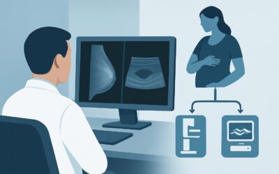
- Mammography screening and Digital breast tomosynthesis (DBT) screening are rated May be appropriate. While they are the workhorses of screening in the general population, their sensitivity is significantly reduced by dense breast tissue. Furthermore, initiating annual screening with an imaging modality that uses ionizing radiation (RRL ☢☢ 0.1-1mSv) at age 25, for example, raises concerns about cumulative radiation exposure over a lifetime.
- US breast is also rated May be appropriate. It does not use radiation and can detect cancers missed by mammography in dense breasts. However, it is highly operator-dependent, has a lower sensitivity than MRI, and is associated with a higher rate of false-positive findings, leading to unnecessary biopsies.
An abbreviated breast MRI protocol is also considered Usually Appropriate. These shorter protocols can improve patient access and reduce costs while maintaining high diagnostic performance for cancer detection, making them an excellent alternative to the full protocol where available. Both MRI options have a radiation level of O (0 mSv), a critical advantage for this young screening population.
Once you’ve decided on MRI breast without and with IV contrast, our protocol guide covers the technique, contrast, and reading principles: MRI Breast With and Without Contrast.
What’s Next After MRI? Downstream Workflow
The results of the screening breast MRI, reported using the Breast Imaging Reporting and Data System (BI-RADS), will dictate the next steps in the patient’s care.
- Negative or Benign Finding (BI-RADS 1 or 2): If the MRI is negative or shows unequivocally benign findings, the patient should be counseled to continue with her annual screening MRI. No further immediate workup is needed.
- Probably Benign Finding (BI-RADS 3): This category is for a finding that has a very high likelihood of being benign (<2% chance of malignancy). The standard recommendation is a short-interval follow-up MRI, typically in 6 months, to ensure stability. This avoids biopsy for many benign lesions while maintaining a high safety margin.
- Suspicious Finding (BI-RADS 4 or 5): These findings warrant a biopsy to establish a definitive diagnosis. The radiologist will recommend the most appropriate biopsy method. Since the finding was detected on MRI, an MRI-guided core needle biopsy is often the necessary next step. If the lesion is also visible on ultrasound, a US-guided biopsy may be performed as it is faster and more accessible.
- Incomplete (BI-RADS 0): This indicates that additional imaging is needed for a complete evaluation. This may involve targeted ultrasound to correlate with an MRI finding or diagnostic mammography if a specific feature needs further characterization.
This structured downstream pathway ensures that positive findings are addressed promptly while minimizing unnecessary interventions for benign or probably benign results.
Pitfalls to Avoid (and When to Get Help)
Navigating screening for this specific patient population requires avoiding several common pitfalls. First, do not use mammography as the sole screening modality; its reduced sensitivity in dense breasts makes it inadequate for this high-risk scenario. Second, remember to time the MRI with the patient’s menstrual cycle—ideally between days 7 and 14—to minimize benign background parenchymal enhancement that can obscure or mimic disease. Third, always screen for contraindications to MRI and gadolinium contrast, including incompatible metallic implants, severe claustrophobia, and impaired renal function (check GFR). If a patient cannot undergo MRI, a consultation with a breast imaging specialist is crucial to determine the best alternative strategy, which typically involves a combination of mammography/tomosynthesis and ultrasound. If any finding is indeterminate or the clinical picture is confusing, escalate the case for discussion at a multidisciplinary breast conference.
Related ACR Topics and Tools
This article focuses on a single, specific clinical scenario. For a comprehensive overview of all patient presentations and risk levels, it is essential to consult the parent topic article and utilize tools that can help you navigate adjacent scenarios.
- For breadth across all scenarios in Female Breast Cancer Screening, see our parent guide: Female Breast Cancer Screening: ACR Appropriateness Decoded.
- Explore other clinical situations using the ACR Appropriateness Criteria Lookup.
- Review technical specifications for recommended studies in our Imaging Protocol Library.
- Discuss cumulative exposure with patients using the Radiation Dose Calculator.
Frequently Asked Questions
Why not just start with mammography for a high-risk woman under 30?
Mammography is rated ‘May be appropriate’ but not ‘Usually Appropriate’ for two main reasons in this group. First, women under 30 almost universally have extremely dense breast tissue, which lowers the sensitivity of mammography, as dense tissue can mask cancers. Second, starting annual radiation-based imaging at such a young age (e.g., 25) raises concerns about cumulative radiation dose over decades of screening. MRI avoids both of these issues.
What specifically defines ‘high risk’ for a woman younger than 30?
A woman under 30 is considered high risk if she has a calculated lifetime risk of breast cancer of 20-25% or more, a known pathogenic variant in a gene like BRCA1 or BRCA2 (or an untested first-degree relative with one), a history of chest radiation between ages 10 and 30, or a diagnosis of a predisposing genetic syndrome like Li-Fraumeni.
Is an abbreviated breast MRI protocol as good as a full protocol?
Yes, for screening purposes in this scenario, the ACR rates an abbreviated breast MRI as ‘Usually Appropriate,’ equivalent to the full protocol. Abbreviated protocols use fewer sequences, significantly shortening the scan time and reducing cost while maintaining very high sensitivity for detecting clinically significant cancers. Their adoption can improve access to MRI screening.
How often should a high-risk woman under 30 be screened?
Screening with breast MRI should be performed annually. The American Cancer Society recommends starting MRI screening at age 25 to 30 for most high-risk women, depending on the specific risk factor.
What is the best screening strategy if my high-risk patient cannot have an MRI?
If a patient has a contraindication to MRI (e.g., a non-compatible implanted device, severe claustrophobia, or gadolinium allergy), the decision should be made in consultation with a breast imaging specialist. The ACR rates both digital breast tomosynthesis (DBT) and breast ultrasound as ‘May be appropriate.’ Often, a combination of both annual DBT and annual ultrasound is used as the best available alternative to MRI.
Reviewed by Pouyan Golshani, MD, Interventional Radiologist — May 30, 2026