Which Imaging Is Best for Local Staging of a Pancreatic Neuroendocrine Tumor?
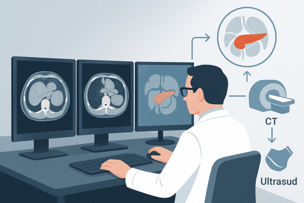
A 58-year-old patient presents to your clinic after an abdominal ultrasound for unrelated symptoms incidentally revealed a 2.5 cm solid mass in the tail of the pancreas. A subsequent endoscopic ultrasound-guided biopsy confirms a well-differentiated neuroendocrine tumor. The patient is otherwise healthy, and the immediate clinical question is surgical planning. To determine resectability, you need to precisely define the tumor’s local extent, its relationship to critical blood vessels, and the status of regional lymph nodes. This article details the clinical workflow for this specific scenario, guiding you to the most appropriate initial imaging study. Based on the American College of Radiology (ACR) Appropriateness Criteria, the recommended first step, CT abdomen and pelvis with IV contrast, is rated Usually appropriate.
Who Fits This Clinical Scenario?
This guidance applies to adult patients with a newly diagnosed or highly suspected pancreatic neuroendocrine tumor (pNET) where the primary clinical goal is local staging. The key questions to be answered are:
- What is the precise size and location of the primary tumor?
- Is there invasion into adjacent structures like the duodenum, stomach, or common bile duct?
- What is the tumor’s relationship to major blood vessels, such as the superior mesenteric artery (SMA), celiac axis, and the portal/superior mesenteric vein (SMV) confluence?
- Are regional lymph nodes enlarged or suspicious for involvement?
This workflow is specifically for defining the T-stage (tumor) and N-stage (nodes) to determine if the patient is a candidate for surgical resection.
This article does not apply to patients in the following situations, which represent distinct clinical scenarios:
- Primary search for distant metastases: If the main goal is to find disease outside the pancreas (e.g., in the liver or bones), the workup follows the “Evaluation for metastatic disease” pathway, which often prioritizes different imaging modalities.
- Post-treatment surveillance: This guidance is not for routine follow-up imaging in patients who have already undergone surgical resection and have no signs of recurrence.
- Monitoring known, untreated disease: Patients with known, unresected pNETs who are undergoing observation or medical therapy follow a separate surveillance imaging schedule.
What Diagnoses Are You Working Up in This Scenario?
While the diagnosis of a pancreatic neuroendocrine tumor is often established or strongly suspected before this stage, the imaging workup is designed to differentiate between stages of local disease, which dictates the entire treatment plan. The “differential” in this context is one of staging, not of initial diagnosis.
Resectable Disease (T1-T3, N0-N1, M0): This is the ideal finding. The imaging would show a tumor confined to the pancreas or with minimal local extension, without encasement of major arteries or veins. The relationship with the portal vein/SMV confluence is critical; clear separation or minimal abutment (<180 degrees of circumference) suggests a high likelihood of successful surgical removal.
Borderline Resectable Disease: This category involves tumors that have more extensive contact with nearby blood vessels. Imaging may show abutment of the SMA, short-segment encasement of the hepatic artery, or abutment or encasement of the portal vein/SMV that may still be amenable to surgical resection and vascular reconstruction. Identifying this stage is crucial for referral to a high-volume pancreatic surgery center.
Locally Advanced / Unresectable Disease: This is a key diagnosis to make non-invasively to avoid unnecessary surgery. Imaging findings would include encasement (>180 degrees of circumference) of the SMA or celiac axis, or unreconstructible occlusion of the portal vein/SMV. The study also assesses for extensive regional lymphadenopathy that may preclude a curative surgical approach.
Why Is CT Abdomen and Pelvis with IV Contrast the Recommended Study for This Presentation?
For the initial local staging of a pancreatic neuroendocrine tumor, a multiphasic contrast-enhanced CT provides the essential anatomical detail required for surgical planning, earning it a rating of Usually appropriate from the ACR.
The rationale is grounded in the typical biology of pNETs. Most well-differentiated pNETs are hypervascular, meaning they have a rich blood supply. This characteristic makes them highly conspicuous on specific phases of a CT scan after intravenous contrast administration. A dedicated pancreas protocol CT, which includes late arterial and portal venous phases, is optimized to exploit this feature. The tumor often appears as a brightly enhancing mass against the normally enhancing pancreatic tissue, allowing for clear delineation of its margins.
Furthermore, CT offers superb spatial resolution for evaluating the critical interface between the tumor and adjacent vascular structures. It can accurately depict abutment versus encasement of the SMA and portal vein, which is the primary determinant of resectability. It also provides a comprehensive survey of the entire abdomen for assessing regional lymph nodes and screening the liver for synchronous metastases, even if that is not the primary question.
Comparing the Alternatives:
- MRI abdomen without and with IV contrast: This study is rated May be appropriate (Disagreement). While MRI can be excellent for problem-solving, particularly for characterizing liver lesions, it is often considered a secondary tool for local pNET staging. CT is generally faster, more widely available, and provides superior visualization of vascular anatomy for many radiologists, which is the key question in this scenario.
- US abdomen endoscopic (EUS): Rated May be appropriate, EUS offers unparalleled high-resolution imaging of the pancreas and adjacent nodes. It is excellent for detecting small tumors and guiding biopsy. However, its field of view is limited, it is operator-dependent, and it cannot provide the global overview of vascular structures and distant organs that CT can, making it complementary rather than a primary staging tool for surgical planning.
- CT abdomen and pelvis without IV contrast: This is rated Usually not appropriate. Without IV contrast, the tumor may be isodense to the surrounding pancreas and therefore invisible. Contrast is essential for both tumor detection and assessment of vascular involvement.
The recommended CT study involves a radiation dose of ☢☢☢ 1-10 mSv. While a full multiphasic study (CT abdomen and pelvis without and with IV contrast) is also Usually appropriate, it carries a higher radiation dose (☢☢☢☢ 10-30 mSv) and may not be necessary if a well-timed single post-contrast acquisition is performed.
Once you’ve decided on the study, our protocol guide covers the technique, contrast, and reading principles: CT Chest/Abdomen/Pelvis with IV Contrast.
What’s Next After CT abdomen and pelvis with IV contrast? Downstream Workflow
The results of the staging CT will direct the patient to one of several distinct clinical pathways. A multidisciplinary tumor board discussion involving surgeons, medical oncologists, and radiologists is standard practice for finalizing the treatment plan.
- If the CT shows clearly resectable disease: The patient should be referred for surgical consultation at a center with expertise in pancreatic surgery. The specific operation (e.g., Whipple procedure, distal pancreatectomy, or enucleation) will depend on the tumor’s location and size.
- If the CT shows borderline resectable or locally advanced disease: The next step is typically a multidisciplinary review. This may lead to consideration of neoadjuvant therapy (treatment before surgery) to shrink the tumor and pull it away from blood vessels. This could involve medical therapies or Peptide Receptor Radionuclide Therapy (PRRT) if the tumor is somatostatin receptor-positive. A
DOTATATE PET/CT skull base to mid-thigh, rated Usually appropriate, is often the next test to confirm somatostatin receptor expression and guide these therapies. - If the CT is indeterminate or equivocal: For instance, if a liver lesion is seen but is too small to characterize, or the relationship to a vein is unclear, a follow-up study is warranted.
MRI abdomen without and with IV contrastis an excellent problem-solver for the liver. EUS can provide a second look at the tumor-vessel interface with high resolution.
Pitfalls to Avoid (and When to Get Help)
Navigating the local staging of a pNET requires attention to detail to avoid common errors that can impact patient care.
- Pitfall 1: Ordering the wrong CT protocol. A standard “CT Abdomen/Pelvis with contrast” may only include a single portal venous phase. This can miss a hypervascular pNET. Always specify a “pancreas protocol” or multiphasic CT to ensure a late arterial phase is acquired.
- Pitfall 2: Underestimating vascular involvement. The distinction between tumor abutment (<180 degrees) and encasement (>180 degrees) of a vessel is subtle but has major implications for resectability. If there is any ambiguity, the case should be reviewed by an experienced abdominal radiologist and discussed at a multidisciplinary tumor board.
- Pitfall 3: Focusing only on the primary tumor. While the task is local staging, the CT provides a comprehensive view of the abdomen. The liver must be carefully scrutinized for any suspicious lesions, as the presence of metastases would fundamentally change the patient’s stage and treatment plan, shifting them to a different clinical pathway.
If the CT report indicates definite vascular encasement or findings suggestive of metastatic disease, escalate the case promptly to a medical oncologist and a multidisciplinary tumor board before proceeding with surgical consultation.
Related ACR Topics and Tools
This article is a deep dive into one specific clinical scenario. For a broader view of imaging across all presentations of this condition, or for details on other clinical questions, the following resources are available.
- For breadth across all scenarios in Staging and Follow-up of Pancreatic Neuroendocrine Tumors, see our parent guide: Staging and Follow-up of Pancreatic Neuroendocrine Tumors: ACR Appropriateness Decoded.
- ACR Appropriateness Criteria Lookup — for adjacent scenarios
- Imaging Protocol Library — for technique on the recommended study
- Radiation Dose Calculator — for cumulative dose conversations
Frequently Asked Questions
Why is DOTATATE PET/CT also rated ‘Usually Appropriate’ for local staging?
DOTATATE PET/CT is excellent for identifying somatostatin receptor (SSTR)-positive disease, which includes most well-differentiated pNETs. It is highly sensitive for both the primary tumor and nodal or distant metastases. While it is rated ‘Usually Appropriate,’ multiphasic CT is often the initial test because it provides superior anatomical detail of vascular structures, which is the most critical question for determining surgical resectability. DOTATATE PET/CT is often used next, especially if metastatic disease is suspected or if PRRT is being considered.
If my patient has a contrast allergy, is MRI the best alternative?
Yes, for a patient with a severe iodinated contrast allergy, MRI is a strong alternative. ‘MRI abdomen without and with IV contrast’ (using a gadolinium-based agent) is rated ‘May be appropriate (Disagreement)’. It can provide excellent soft tissue contrast and, with specific sequences, can evaluate vascular involvement. Pre-medication for a CT is also an option for many patients with a history of mild to moderate reactions.
What if the pNET was found incidentally on a non-contrast CT?
A non-contrast CT is considered insufficient for staging. If a pNET is suspected on a non-contrast study (e.g., as a calcified or partially cystic mass), the next step should be a dedicated, contrast-enhanced pancreas protocol CT or MRI to properly delineate the tumor and its relationship to surrounding structures as outlined in this guide.
Does tumor functionality (e.g., an insulinoma) change the initial local staging imaging choice?
No, the choice of imaging for initial *local anatomical staging* remains the same regardless of whether the tumor is functional or non-functional. The goal is to define the anatomy for surgical planning. While functional status is critical for patient management, the primary role of CT or MRI in this scenario is to answer the ‘can we resect this?’ question based on anatomical relationships.
Should the CT include the chest?
While the ACR scenario specifies ‘CT abdomen and pelvis,’ many institutional protocols for oncologic staging will include the chest to provide a complete baseline assessment for metastatic disease, as the lungs are a potential site. This is a reasonable consideration and should be discussed with the consulting radiologist, though the primary focus for local staging remains the detailed multiphasic imaging of the pancreas.
Reviewed by Pouyan Golshani, MD, Interventional Radiologist — May 30, 2026