Which Imaging Is Best for Uterine Fibroid Treatment Planning? An ACR Workflow
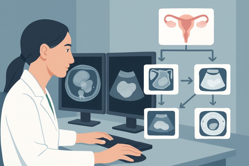
A 42-year-old woman with a known history of uterine fibroids returns to your clinic. Her symptoms of heavy menstrual bleeding and pelvic pressure have worsened, and she is now ready to discuss definitive treatment options, including uterine artery embolization (UAE) and myomectomy. Before you can refer her to interventional radiology or gynecology for a procedure, you need to provide a detailed map of her fibroids—their number, size, location, and blood supply. This article details the ACR-guided imaging workflow for this specific clinical decision: selecting the initial imaging for treatment planning in a patient with known fibroids. For this scenario, the American College of Radiology rates US duplex Doppler pelvis as Usually Appropriate.
## Who Fits This Clinical Scenario for Fibroid Treatment Planning?
This guidance applies specifically to patients with an established diagnosis of uterine leiomyomas (fibroids) who are actively considering procedural intervention. The primary goal of imaging in this context is not diagnosis, but pre-procedural characterization and mapping to determine treatment eligibility and approach.
Inclusion criteria for this workflow:
- A confirmed, pre-existing diagnosis of uterine fibroids.
- The patient is symptomatic and seeking treatment (e.g., myomectomy, hysterectomy, uterine artery embolization, radiofrequency ablation).
- The clinical question is focused on defining fibroid anatomy and vascularity to guide the choice and execution of a specific therapy.
This workflow does NOT apply to:
- Patients with suspected but unconfirmed fibroids: This presentation falls under a different clinical variant, “Clinically suspected fibroids. Initial imaging,” which focuses on initial diagnosis rather than pre-operative planning.
- Patients requiring follow-up after treatment: Imaging to assess treatment response or for surveillance is covered by the “Known fibroids. Surveillance or posttreatment imaging” scenario, which has different considerations.
- Patients presenting with acute, severe symptoms: An acute presentation, such as suspected fibroid torsion or severe hemorrhage, may require a more emergent and potentially different imaging protocol to rule out urgent complications.
## What Are You Characterizing for Treatment Planning?
Because the diagnosis is already known, the imaging “workup” shifts from detection to detailed characterization. The goal is to answer specific questions that directly influence which treatments are feasible and safe.
Fibroid Mapping (Number, Size, and Location)
This is the foundational task. The imaging report must clearly delineate each significant fibroid’s location relative to the uterine layers. Is it submucosal (impinging on the endometrial cavity, often causing heavy bleeding), intramural (within the uterine wall), or subserosal (projecting from the outer surface)? A pedunculated fibroid (attached by a stalk) carries a risk of torsion. This mapping is critical; a submucosal fibroid may be amenable to a minimally invasive hysteroscopic myomectomy, whereas a large intramural fibroid may require a laparoscopic/robotic or open myomectomy or UAE.
Assessing Vascularity
For treatments like uterine artery embolization, understanding the fibroid’s blood supply is paramount. Doppler ultrasound helps identify the feeding vessels and assess the degree of vascularity within the fibroid itself. This information confirms that the fibroid is a viable target for embolization and helps interventional radiologists plan their approach.
Evaluating for Alternative Pathology
Symptoms attributed to fibroids can sometimes be caused or exacerbated by co-existing conditions. Imaging helps differentiate or confirm the presence of adenomyosis, a condition where endometrial tissue grows into the uterine wall, which can present similarly but requires a different treatment strategy. The workup should also confidently exclude other adnexal pathology, such as ovarian cysts or masses, as the source of the patient’s symptoms.
Screening for Features Suggestive of Malignancy
While exceedingly rare, the possibility of a uterine leiomyosarcoma must be considered. Imaging cannot definitively diagnose malignancy, but certain features on MRI, such as rapid growth, irregular infiltrative margins, or areas of central necrosis, can raise suspicion. Identifying these red flags is crucial, as it may preclude procedures like morcellation during myomectomy, which can disseminate malignant cells.
## Why Is Pelvic Ultrasound with Doppler the Recommended First Step for Treatment Planning?
The ACR designates multiple modalities as Usually Appropriate for this scenario, but pelvic ultrasound with Doppler is an excellent, accessible, and often sufficient first step for initial treatment planning.
The complete examination typically includes both transabdominal and transvaginal approaches. The transabdominal view provides a broad overview of the pelvis, which is essential for assessing very large, superiorly located, or exophytic fibroids that may be out of reach of the transvaginal probe. The transvaginal portion offers superior spatial resolution, providing detailed characterization of the endometrium, myometrium, and smaller fibroids.
The “duplex Doppler” component is what makes this study particularly valuable for treatment planning. Color and spectral Doppler imaging assesses blood flow within the myometrium and the fibroids themselves. This is critical for confirming vascular supply ahead of a planned uterine artery embolization.
Comparison to Other Modalities:
- MRI Pelvis without and with IV Contrast is also rated Usually Appropriate and is considered the gold standard for comprehensive fibroid mapping. It provides superior soft tissue contrast, allowing for precise localization of all fibroids, clear differentiation from adenomyosis, and detailed assessment of internal characteristics. For complex cases involving numerous fibroids (a “bag of oranges” uterus) or when planning a difficult myomectomy, many specialists will proceed directly to MRI. The choice between initial US and MRI often depends on the complexity of the case and the specific intervention planned.
- MRI Pelvis without IV Contrast is rated May be appropriate. While it still provides excellent anatomical detail for mapping fibroid number and location, the lack of gadolinium-based contrast limits the assessment of fibroid vascularity and viability, which is important information for many treatment pathways.
- CT Pelvis (with or without contrast) is rated Usually not appropriate. CT offers poor soft-tissue resolution for differentiating myometrial pathology compared to US or MRI. More importantly, it exposes the patient, who is often of reproductive age, to unnecessary ionizing radiation (ACR Relative Radiation Level ☢☢☢ to ☢☢☢☢) without providing the detailed characterization needed for procedural planning.
## What Happens After the Initial Ultrasound? Downstream Workflow
The results of the initial ultrasound will guide the next steps in a logical, tiered fashion.
- If the ultrasound is sufficient for planning: For a patient with a few, clearly delineated fibroids who is considering a straightforward myomectomy or UAE, a high-quality ultrasound may provide all the necessary information. The patient can be referred directly to the appropriate specialist (gynecology or interventional radiology) with the imaging report.
- If the ultrasound is indeterminate or shows high complexity: If the ultrasound reveals a very large number of fibroids, making an accurate count difficult, or if there is a strong suspicion of co-existing adenomyosis, the next step is to obtain the more definitive study. An MRI of the pelvis with and without IV contrast is the logical next step to create a precise surgical or procedural map.
- If the ultrasound is negative for significant fibroids: In the rare case that a patient with a “known” history of fibroids has an ultrasound that does not demonstrate a clear cause for their symptoms, the diagnosis should be reconsidered. The workup may need to shift toward evaluating for other causes of abnormal uterine bleeding or pelvic pain, such as adenomyosis or endometrial pathology.
## Pitfalls to Avoid (and When to Get Help)
- Underestimating Fibroid Burden on US: Ultrasound is operator-dependent, and in a uterus with many fibroids, it can be difficult to count and measure each one accurately. Be aware of this limitation and have a low threshold to proceed to MRI for complex cases.
- Mistaking Adenomyosis for Fibroids: While classic features exist for both, diffuse adenomyosis or an adenomyoma can sometimes be difficult to distinguish from a leiomyoma on ultrasound. If the imaging pattern is atypical, MRI is the superior problem-solving tool.
- Ignoring the Endometrial Stripe: Always ensure the report comments on the endometrial lining. A submucosal fibroid can distort the endometrium, and any associated endometrial thickening or irregularity may warrant further investigation (e.g., sonohysterography or biopsy) to rule out concurrent pathology.
If the clinical picture and imaging findings are discordant, or if there is any suspicion of malignancy, consultation with a gynecologic oncologist or a radiologist specializing in pelvic imaging is warranted before proceeding with any intervention.
## Related ACR Topics and Tools
This article covers one specific clinical scenario. For a broader view of all fibroid-related imaging decisions or to explore the technical details of the recommended studies, the following resources are available. For breadth across all scenarios in Fibroids, see our parent guide: Fibroids: ACR Appropriateness Decoded.
- ACR Appropriateness Criteria Lookup — For exploring adjacent clinical scenarios and alternative imaging guidelines.
- Imaging Protocol Library — For technical specifications on how imaging studies like pelvic MRI and ultrasound are performed.
- Radiation Dose Calculator — For discussing cumulative radiation exposure with patients when considering studies like CT.
Frequently Asked Questions
Is an MRI always necessary before uterine artery embolization (UAE)?
Not always, but it is very common and often preferred. While a high-quality duplex Doppler ultrasound can provide essential information on uterine artery anatomy and fibroid vascularity, MRI is superior for confirming that symptoms are truly from fibroids (vs. adenomyosis), mapping all existing fibroids, and identifying any contraindications to the procedure. Many interventional radiology practices consider MRI a prerequisite for UAE.
Why is CT rated ‘Usually not appropriate’ for fibroid planning?
CT is not recommended for two main reasons. First, it uses ionizing radiation, which is a key consideration in the typically reproductive-age female population being treated for fibroids. Second, its ability to differentiate soft tissues within the pelvis (like distinguishing fibroids from normal myometrium or adenomyosis) is significantly inferior to both ultrasound and MRI. It does not provide the detailed anatomical map needed for effective treatment planning.
If a patient has an IUD, can she still get a transvaginal ultrasound and MRI?
Yes. Most modern intrauterine devices (IUDs), including hormonal and copper IUDs, are safe for both transvaginal ultrasound and MRI (typically up to 3 Tesla). The IUD will be visible on imaging and can create some local artifact, but it does not prevent a diagnostic quality examination for fibroid mapping.
What is the role of sonohysterography in fibroid treatment planning?
Sonohysterography (saline-infusion sonography) is a specialized ultrasound technique where sterile saline is infused into the endometrial cavity. It is an excellent problem-solving tool specifically for evaluating the relationship of fibroids to the endometrium. It is the best modality to definitively confirm if a fibroid is submucosal and to what degree it projects into the cavity, which is critical information when planning a hysteroscopic myomectomy.
Does the phase of the menstrual cycle matter when scheduling the ultrasound or MRI?
For general fibroid mapping, the timing within the menstrual cycle is not critical. However, if there is a need to evaluate the endometrium with high precision, imaging is often best performed in the early proliferative phase (e.g., days 4-10 of the cycle), when the endometrium is at its thinnest, making it easier to detect and characterize intracavitary pathology like submucosal fibroids or polyps.
Reviewed by Pouyan Golshani, MD, Interventional Radiologist — May 29, 2026