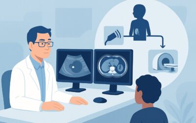
Which Imaging Study Is Best for a Child with Refractory Epilepsy?

A 9-year-old patient is in your pediatric neurology clinic for follow-up. Their seizures began at age 5 and have become progressively more difficult to control, now occurring weekly despite trials of three different anti-epileptic medications. The family is seeking answers, and you are beginning to consider whether the child might be a candidate for epilepsy surgery. The critical first step in that evaluation is identifying a potential structural cause. What is the optimal imaging study to order for a child with intractable seizures to guide this complex decision-making process?
According to the American College of Radiology (ACR) Appropriateness Criteria, for a child with intractable seizures or refractory epilepsy, an MRI head without IV contrast is the imaging study that is Usually Appropriate.
Which Children Fit the Refractory Epilepsy Imaging Scenario?
This clinical workflow is specifically for children aged 1 month to 17 years who have been diagnosed with intractable or refractory epilepsy. This diagnosis is generally made when seizures persist despite adequate trials of two or more tolerated and appropriately chosen anti-epileptic drug (AED) schedules, whether used in combination or as monotherapy. The key element is the failure of medical management, which shifts the clinical focus from seizure control to identifying an underlying, potentially resectable, structural cause.
It is crucial to distinguish this scenario from other common pediatric seizure presentations that follow different imaging pathways:
- Initial Seizure Workup: This guidance does not apply to the first-time workup of a focal or primary generalized seizure in a neurologically normal child.
- Febrile Seizures: Children presenting with simple or complex febrile seizures have distinct evaluation criteria and typically do not require advanced imaging.
- Neonatal Seizures: Seizures in the first month of life (0 to 29 days) represent a unique clinical entity with a different differential diagnosis and imaging approach.
- Post-Traumatic Seizures: When seizures occur after head trauma, the imaging priority is to assess for acute injury like hemorrhage or contusion, often involving different modalities.
This article is exclusively for the non-emergent, diagnostic evaluation of a child whose seizures have proven resistant to standard medical therapy.
What Diagnoses Are You Working Up in a Child with Intractable Seizures?
When seizures are refractory to medication, the likelihood of an underlying structural brain abnormality increases significantly. The imaging workup is designed to identify these epileptogenic lesions, many of which are subtle. The primary differential diagnoses include:
Focal Cortical Dysplasia (FCD): FCD is a malformation of cortical development and one of the most common causes of intractable epilepsy in children. These areas of disorganized brain cortex are highly epileptogenic. They can be subtle and are often characterized by abnormalities in cortical thickness, blurring of the gray-white matter junction, and abnormal signal on specific MRI sequences. High-resolution imaging is essential for their detection.
Mesial Temporal Sclerosis (MTS): Also known as hippocampal sclerosis, MTS is the most frequent cause of refractory focal epilepsy in adults but is also a key consideration in adolescents. It involves neuronal loss and scarring (gliosis) in the hippocampus. Key imaging findings include hippocampal volume loss and increased signal on T2-weighted and FLAIR sequences.
Low-Grade Neoplasms: Slow-growing tumors are another important cause of refractory seizures in the pediatric population. Tumors such as gangliogliomas and dysembryoplastic neuroepithelial tumors (DNETs) are often located in the temporal lobe, present with seizures as the primary symptom, and may be amenable to surgical resection.
Vascular Malformations: Lesions like cavernous malformations (cavernomas) can cause seizures due to chronic irritation from small hemorrhages and hemosiderin deposition in the adjacent brain tissue. While less common, arteriovenous malformations (AVMs) can also be a source of seizures.
Post-Insult Gliosis: Brain scarring resulting from a past event—such as a perinatal stroke, infection (encephalitis), or significant head trauma—can create a persistent focus for seizure activity. Imaging helps identify the location and extent of such scarring.
Why Is Non-Contrast MRI the Recommended Study for Refractory Epilepsy?
The ACR designates MRI head without IV contrast as Usually Appropriate because it provides the best balance of diagnostic yield and safety for identifying the structural causes of refractory epilepsy.
The primary strength of Magnetic Resonance Imaging (MRI) is its superior soft tissue contrast and spatial resolution. This allows for detailed visualization of the brain’s anatomy, including the subtle distinctions between gray and white matter and the intricate structure of the cerebral cortex and hippocampus. This capability is critical for detecting the subtle findings of Focal Cortical Dysplasia and Mesial Temporal Sclerosis, which are often completely invisible on other imaging modalities like CT.
For this initial structural evaluation, intravenous contrast is typically not required. The key diagnostic features of FCD and MTS are best seen on non-contrast sequences. Therefore, performing the study without contrast avoids unnecessary exposure to gadolinium-based contrast agents in the pediatric population.
Why Other Studies Are Rated Lower
- CT Head (all variants): Rated Usually not appropriate. Computed Tomography (CT) lacks the sensitivity to detect the most common causes of refractory epilepsy, such as FCD and MTS. Furthermore, CT exposes the child to ionizing radiation (pediatric dose ☢☢☢ 0.3-3 mSv for a non-contrast study), which should be avoided when a non-radiation modality offers superior diagnostic information.
- MRI Head without and with IV contrast: Rated May be appropriate (Disagreement). While contrast can help characterize a suspected tumor or vascular lesion, the ACR panel notes disagreement on its routine use. Many epilepsy centers advocate for a high-quality non-contrast epilepsy protocol first, reserving contrast for cases where a specific finding on the initial images warrants further characterization.
When ordering, it is essential to specify an “MRI Brain Epilepsy Protocol.” This communicates to the radiology department that specialized, high-resolution, thin-slice sequences (such as T1, T2, and FLAIR) are needed. These sequences are optimized to detect the subtle epileptogenic lesions that a standard brain MRI protocol might miss.
What Are the Next Steps After the Initial MRI Results?
The results of the epilepsy protocol MRI are a critical branch point in the patient’s management. The downstream workflow depends directly on the findings.
If the MRI is positive for a resectable lesion (e.g., FCD, tumor, MTS): This is a significant finding that makes the patient a potential candidate for epilepsy surgery. The next step is typically a multidisciplinary epilepsy surgery conference. This team—including pediatric neurologists, neurosurgeons, neuroradiologists, and neuropsychologists—will review the case. Further evaluation may include advanced functional imaging like FDG-PET or SPECT to confirm the lesion is the source of the seizures, as well as video-EEG monitoring to precisely map the seizure onset zone relative to the lesion.
If the MRI is negative or non-localizing: A “normal” MRI does not rule out a structural cause, as some lesions can be “MRI-negative.” In this situation, the workup may proceed to functional imaging studies. Both FDG-PET/CT brain and SPECT brain perfusion are rated May be appropriate. These studies assess brain metabolism and blood flow, respectively, and can identify an abnormal area (hypometabolism on interictal PET, hyperperfusion on ictal SPECT) that may represent the seizure focus, even without a clear structural correlate on MRI.
If the MRI is indeterminate: Ambiguous findings may require further characterization. This could involve a follow-up MRI with contrast, a higher field-strength MRI (e.g., 3T or 7T if available), or correlation with functional imaging and EEG data to determine the clinical significance of the finding.
Pitfalls to Avoid (and When to Get Help)
Navigating the workup for pediatric refractory epilepsy requires careful attention to detail. Here are a few common pitfalls to avoid:
- Ordering a standard brain MRI: A routine MRI protocol may lack the specific thin-slice sequences and angulations needed to detect subtle cortical dysplasias or hippocampal sclerosis. Always specify “epilepsy protocol.”
- Accepting a “normal” report at face value: If clinical suspicion for a structural lesion is high, a negative MRI report warrants a second look. Consider having the images reviewed by a neuroradiologist with specific expertise in epilepsy imaging.
- Delaying the workup: Ongoing, uncontrolled seizures can have significant neurodevelopmental consequences. Once a child meets the criteria for refractory epilepsy, a timely and thorough imaging evaluation is crucial.
- Ignoring functional data: MRI provides anatomical information. Always correlate imaging findings with the patient’s seizure semiology and EEG results to confirm that a detected lesion is the true epileptogenic zone.
If the initial non-contrast MRI is negative but the clinical and EEG data strongly point to a focal seizure onset, escalate the case to a comprehensive epilepsy center for consideration of advanced imaging and multidisciplinary evaluation.
Related ACR Topics and Tools
The ACR Appropriateness Criteria are a valuable resource for guiding complex imaging decisions. For a comprehensive overview of all pediatric seizure scenarios, from neonatal to post-traumatic, please see our parent guide. Additional tools can help you apply these guidelines in your practice.
- For breadth across all scenarios in Seizures-Child, see our parent guide: Seizures-Child: ACR Appropriateness Decoded.
- To explore other clinical scenarios and their corresponding imaging recommendations, visit the Imaging Appropriateness Selector.
- For technical details on performing the recommended study, consult the Imaging Protocol Library.
- To discuss radiation exposure from alternative studies like CT or PET/CT with families, use the Radiation Dose Calculator.
Frequently Asked Questions
Why is a non-contrast MRI preferred over one with contrast for refractory epilepsy?
A non-contrast MRI is preferred because the most common structural causes of refractory epilepsy in children, such as focal cortical dysplasia (FCD) and mesial temporal sclerosis (MTS), are best visualized on high-resolution non-contrast sequences (like T1, T2, and FLAIR). Intravenous contrast does not typically improve the detection of these specific conditions and adds unnecessary exposure to a gadolinium-based agent. Contrast is reserved for cases where a tumor or vascular malformation is suspected based on the initial non-contrast images.
What should I do if the epilepsy protocol MRI is reported as normal?
A ‘normal’ or ‘negative’ MRI does not end the workup. If clinical and EEG data strongly suggest a focal seizure origin, the next step is often functional imaging. Studies like FDG-PET (which shows metabolism) or SPECT (which shows blood flow) are rated ‘May be appropriate’ and can help localize a seizure focus even when the brain structure appears normal on MRI. Consultation with a comprehensive epilepsy center is highly recommended.
Is CT ever appropriate for evaluating a child with intractable seizures?
For the specific workup of intractable epilepsy, CT is rated ‘Usually not appropriate’ by the ACR. Its poor soft tissue contrast makes it insensitive for detecting key pathologies like cortical dysplasia. CT is primarily used in emergent settings to rule out acute processes like hemorrhage or hydrocephalus, not for the detailed structural analysis required in refractory epilepsy.
How does the imaging workup differ for a child’s first-time seizure versus refractory seizures?
The workup for a first-time, unprovoked seizure in a neurologically normal child may not always require immediate imaging, and the decision is based on clinical factors. The workup for refractory epilepsy, however, is fundamentally different. Its primary goal is to actively search for a structural, and potentially surgically correctable, cause for the medically uncontrollable seizures, making high-resolution MRI an essential diagnostic step.
What is the role of FDG-PET or SPECT in this scenario?
FDG-PET and SPECT are functional imaging studies rated ‘May be appropriate.’ They are most valuable when the structural MRI is negative or the findings are ambiguous. An interictal (between seizures) FDG-PET scan can show a region of decreased metabolism, while an ictal (during a seizure) SPECT scan can show a region of increased blood flow. These findings can pinpoint the seizure focus and guide further treatment, including potential surgery.
Reviewed by Pouyan Golshani, MD, Interventional Radiologist — May 26, 2026