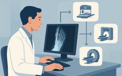
Which Imaging Study Is Best for Suspected Foot Osteomyelitis with Metal Hardware?
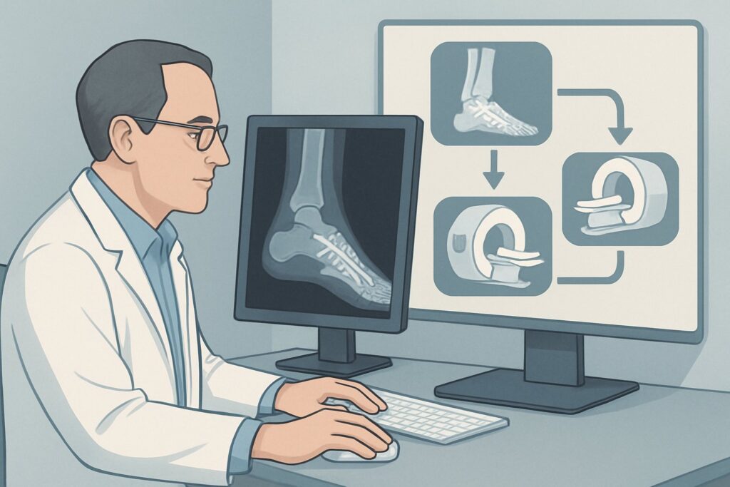
A 64-year-old man with type 2 diabetes and a history of Charcot foot reconstruction presents with a non-healing ulcer over his midfoot, directly overlying a surgical plate and screws. He has localized erythema and warmth, and his C-reactive protein is elevated. You obtain plain radiographs, which show the hardware is intact but are otherwise indeterminate for acute osteomyelitis due to post-surgical changes. You are now faced with a critical decision: which advanced imaging study can accurately diagnose or exclude bone infection in the presence of significant metal artifact? This article provides a step-by-step clinical workflow for this specific, challenging scenario. According to the American College of Radiology (ACR) Appropriateness Criteria, the next step, MRI of the foot without and with IV contrast, is rated Usually Appropriate.
Who Fits This Clinical Scenario?
This guidance is for a specific patient population: an adult with diabetes mellitus who is suspected of having osteomyelitis of the foot, but with two key complicating factors. First, the patient has indwelling metal instrumentation from a prior surgery (e.g., plates, screws, or other internal fixation devices). Second, initial radiographs have already been performed and were either negative or indeterminate for osteomyelitis. This workflow is designed for the “what next?” moment after an unrevealing X-ray.
This article does not apply to several similar-but-distinct clinical situations:
- Patients without metal hardware: If the patient has suspected diabetic foot osteomyelitis and a negative radiograph but no metal instrumentation, the diagnostic considerations are different, as artifact is not a primary concern.
- Initial imaging workup: This guide assumes radiographs have already been performed. For guidance on the very first imaging study to order, a different workflow applies.
- Clearly positive radiographs: If initial X-rays are definitive for osteomyelitis, the imaging goal shifts from diagnosis to pre-procedural planning, which follows a separate ACR variant.
Correctly identifying your patient’s scenario is crucial for selecting the most effective and appropriate imaging test.
What Diagnoses Are You Working Up in This Scenario?
When a patient with diabetes and foot hardware presents with signs of infection, the differential diagnosis is narrow but has significant management implications. The goal of advanced imaging is to distinguish between these possibilities, as their treatments are vastly different.
Osteomyelitis is the primary concern. In diabetic patients, this is most often due to contiguous spread from an overlying skin ulcer. The infection breaches the soft tissue and seeds the bone, potentially involving the hardware itself. Confirming or excluding this diagnosis is paramount, as it dictates the need for long-term antibiotics and often surgical debridement.
Acute or subacute Charcot neuroarthropathy is a key mimic of infection. This non-infectious, inflammatory process of bone and joint destruction can present with erythema, warmth, and swelling, clinically indistinguishable from infection. Since many patients have hardware to stabilize a prior Charcot deformity, a new inflammatory flare can be easily confused with osteomyelitis.
Aseptic hardware loosening or failure is another important consideration. Mechanical stress can cause hardware to loosen from the bone, creating an inflammatory response that can mimic infection. While less common than infection, identifying this directs management toward orthopedic revision rather than antimicrobial therapy.
Finally, a soft tissue abscess or cellulitis without underlying osteomyelitis must be considered. In this case, the infection is confined to the soft tissues and has not yet invaded the bone. Imaging can help delineate the extent of soft tissue involvement and guide potential incision and drainage while sparing the patient a prolonged course of bone-penetrating antibiotics.
Why Is MRI of the Foot Without and With IV Contrast the Recommended Study?
For this complex scenario, Magnetic Resonance Imaging (MRI) of the foot without and with IV contrast is rated Usually Appropriate and is the preferred next step. The primary challenge is obtaining diagnostic-quality images despite the signal distortion caused by metallic hardware. Modern MRI techniques are specifically designed to overcome this.
MRI’s superior soft tissue contrast is its key advantage. It can directly visualize bone marrow edema, the earliest sign of osteomyelitis, long before bony destruction is visible on other modalities. Furthermore, it can clearly delineate sinus tracts extending from a skin ulcer to the bone, identify fluid collections and abscesses, and assess the viability of surrounding tissues. The administration of IV gadolinium-based contrast enhances the conspicuity of abscesses, phlegmon, and non-viable tissue, which is critical for surgical planning.
A common misconception is that metal hardware is an absolute contraindication to MRI. While older techniques were severely limited by artifact, contemporary MRI scanners utilize metal artifact reduction sequences (MARS). When ordering the study, it is crucial to specify the presence and location of hardware so the radiology department can implement these specialized protocols to generate diagnostic images.
Let’s consider the alternatives and why they are rated differently for this specific scenario:
- Nuclear Medicine (3-phase bone scan and WBC scan with SPECT/CT): This combined study is also rated Usually Appropriate. It is a powerful tool for detecting infection, as it relies on physiologic uptake of radiotracers by inflammatory cells. It is less affected by metal artifact than a non-MARS MRI. However, it provides lower spatial resolution, making it harder to define the precise anatomic extent of infection. It also involves significant ionizing radiation (adult RRL ☢☢☢☢, 10-30 mSv) and is more time-consuming. It is an excellent problem-solving tool if MRI is contraindicated or remains indeterminate.
- CT foot with IV contrast: This is rated May be appropriate. CT provides excellent detail of cortical bone and the integrity of the hardware itself. However, it is far less sensitive than MRI for detecting early bone marrow edema and cannot delineate soft tissue infection with the same clarity. It involves a small amount of ionizing radiation (adult RRL ☢, <0.1 mSv).
Given its ability to directly visualize marrow and soft tissue pathology, lack of ionizing radiation (adult RRL O, 0 mSv), and the availability of MARS protocols, MRI with and without contrast provides the most comprehensive diagnostic information in most cases.
What’s Next After MRI? Downstream Workflow
The results of the MRI will guide your subsequent management decisions, steering the patient toward medical, surgical, or conservative pathways.
- If the MRI is positive for osteomyelitis: The report will describe bone marrow signal changes and enhancement consistent with infection, often with an associated abscess or sinus tract. This finding typically necessitates a consultation with infectious disease and podiatric or orthopedic surgery. A bone biopsy, often image-guided, is frequently the next step to obtain microbiology and confirm the diagnosis before initiating a 4-6 week course of targeted antibiotics. Surgical debridement, and potentially hardware removal, may be required.
- If the MRI is negative for osteomyelitis: If the study shows only soft tissue infection (e.g., cellulitis or an abscess) without underlying bone involvement, management can focus on soft tissue drainage and a shorter course of antibiotics. If the MRI instead shows findings of acute Charcot neuroarthropathy without signs of infection, the primary treatment is aggressive offloading (e.g., total contact casting) to prevent further deformity.
- If the MRI is indeterminate: In some cases, even with MARS techniques, artifact from the hardware may obscure the underlying bone and make the study non-diagnostic. In this situation, the next logical step is to pivot to the other Usually Appropriate study: a combined labeled white blood cell and bone marrow scan (often with SPECT/CT), which is less susceptible to metal artifact.
The MRI result is a critical branch point, fundamentally altering the patient’s treatment plan and prognosis.
Pitfalls to Avoid (and When to Get Help)
Navigating this scenario requires careful planning to avoid common errors. First, do not assume MRI is impossible due to hardware; instead, communicate clearly with the radiology department when ordering, specifying the presence of metal so they can apply artifact reduction sequences. Second, avoid ordering a non-contrast MRI if there is no contraindication to gadolinium; contrast is vital for delineating abscesses and assessing tissue viability around the hardware. Third, never rely solely on imaging. The clinical picture, wound appearance, and inflammatory markers are all essential pieces of the puzzle. If the imaging findings are discordant with the clinical presentation, a direct discussion with the radiologist is often the best next step to resolve ambiguity and plan further workup, such as a nuclear medicine study or bone biopsy.
Related ACR Topics and Tools
This article focuses on one specific clinical variant. For a comprehensive overview of imaging for all related scenarios, from initial workup to post-treatment follow-up, please consult the parent topic article. The following GigHz tools can also support your clinical decision-making:
- For breadth across all scenarios in Suspected Osteomyelitis of the Foot in Patients with Diabetes Mellitus, see our parent guide: Suspected Osteomyelitis of the Foot in Patients with Diabetes Mellitus: ACR Appropriateness Decoded.
- ACR Appropriateness Criteria Lookup — for adjacent scenarios
- Imaging Protocol Library — for technique on the recommended study
- Radiation Dose Calculator — for cumulative dose conversations
Frequently Asked Questions
Why not just go straight to a bone biopsy instead of advanced imaging?
A bone biopsy is the gold standard for diagnosing osteomyelitis, but it is an invasive procedure. Advanced imaging like MRI is crucial for pre-procedural planning. It confirms the presence of a bony abnormality, identifies the best and safest location to biopsy, and helps avoid sampling areas of non-diagnostic necrotic tissue or missing an adjacent abscess that also requires drainage.
What if my patient has a contraindication to MRI, like a non-compatible implanted device or severe claustrophobia?
In cases where MRI is contraindicated, the ACR lists a combined 3-phase bone scan and labeled white blood cell (WBC) scan with SPECT or SPECT/CT as another ‘Usually Appropriate’ option. This nuclear medicine study is an excellent alternative for detecting infection and is not limited by most contraindications to MRI.
Is an MRI without contrast sufficient in this scenario?
According to the ACR, an MRI of the foot without IV contrast is also rated ‘Usually Appropriate’. However, an MRI with contrast is generally preferred. The addition of a gadolinium-based contrast agent significantly improves the detection of abscesses, sinus tracts, and non-viable tissue, which is critical information for surgical planning. A non-contrast study may be sufficient to evaluate for bone marrow edema but provides less information about the surrounding soft tissues.
How do metal artifact reduction sequences (MARS) for MRI work?
MARS MRI protocols use a combination of advanced software and hardware techniques to minimize the signal distortion caused by metal. This includes using different magnetic field strengths, modifying pulse sequence parameters (like echo time and bandwidth), and employing specialized image reconstruction algorithms. These adjustments help to reduce the size and intensity of artifacts, allowing the radiologist to better visualize the bone and soft tissues immediately adjacent to the hardware.
If the MRI shows changes of Charcot neuroarthropathy, does that rule out a superimposed infection?
Not necessarily. Differentiating an acute Charcot flare from a superimposed infection is one of the greatest challenges in musculoskeletal imaging. While certain MRI findings are more suggestive of one over the other (e.g., a discrete abscess and sinus tract favor infection), the findings can overlap. In equivocal cases, a labeled WBC scan or a bone biopsy may be necessary to definitively rule out a co-existing infection.
Reviewed by Pouyan Golshani, MD, Interventional Radiologist — May 30, 2026