Which MRI Is Best for Suspected Optic Neuritis? An ACR-Guided Imaging Workflow

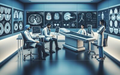
A 28-year-old patient presents to your clinic with three days of worsening, painful vision loss in her right eye. She describes the vision as “blurry” and notes that colors appear washed out. On examination, you find a relative afferent pupillary defect and reduced color vision on the affected side. Your leading diagnosis is optic neuritis, and you know that imaging is the critical next step to confirm the diagnosis and assess for underlying demyelinating disease. The central question is which specific study to order. This article provides a detailed clinical workflow for this exact scenario, grounded in the American College of Radiology (ACR) Appropriateness Criteria. For suspected optic neuritis, the ACR rates MRI head without and with IV contrast as Usually Appropriate.
## Who Fits the Clinical Scenario of Suspected Optic Neuritis?
This guidance applies to patients presenting with symptoms and signs suggestive of acute optic nerve inflammation. The classic presentation involves acute to subacute monocular visual loss that evolves over several days to two weeks, frequently accompanied by pain, especially with eye movement. Key clinical findings that place a patient in this scenario include:
- Reduced visual acuity in one eye.
- A relative afferent pupillary defect (RAPD) on swinging-flashlight test.
- Dyschromatopsia (impaired color vision) that is often disproportionately severe compared to the loss of visual acuity.
- A central or paracentral scotoma on visual field testing.
It is crucial to distinguish this presentation from similar but distinct clinical scenarios that require a different imaging approach. This workflow does not apply if:
- There is a history of significant orbital trauma. A traumatic visual defect requires a workup focused on orbital injury, such as fracture or optic nerve sheath hematoma.
- The primary sign is nontraumatic proptosis or globe displacement. This presentation points toward an orbital mass or thyroid eye disease, which follows a different diagnostic pathway.
- Signs of infection are present. Symptoms like fever, eyelid erythema and edema, and restricted extraocular movements suggest orbital cellulitis, a condition requiring urgent and specific imaging protocols.
## What Diagnoses Are You Working Up with Imaging for Optic Neuritis?
When ordering imaging for suspected optic neuritis, the goal is twofold: to confirm inflammation of the optic nerve and, critically, to search for an underlying systemic cause. The differential diagnosis guides the choice of imaging modality and protocol.
The most common and consequential diagnosis to consider is demyelinating disease, primarily multiple sclerosis (MS). Optic neuritis is the initial presentation of MS in approximately 20% of patients, and an abnormal brain MRI at the time of initial optic neuritis is the single strongest predictor of a future MS diagnosis. Imaging is therefore essential for both diagnosis and prognostication.
Atypical forms of optic neuritis must also be considered, as they carry different prognoses and require different treatments. These include Neuromyelitis Optica Spectrum Disorder (NMOSD), often associated with aquaporin-4 antibodies, and Myelin Oligodendrocyte Glycoprotein (MOG) antibody-associated disease (MOGAD). These conditions can sometimes be distinguished by the pattern of optic nerve involvement on MRI (e.g., longer segments of enhancement, bilateral involvement).
While less common in this acute presentation, a compressive optic neuropathy from a mass such as an optic nerve sheath meningioma or glioma must be excluded. These lesions can sometimes present with progressive visual loss that mimics an inflammatory process.
Finally, various infectious and inflammatory conditions can cause optic neuritis, including sarcoidosis, lupus, syphilis, and Lyme disease. While MRI findings can sometimes be suggestive, these diagnoses are typically confirmed with serologic testing and clinical correlation.
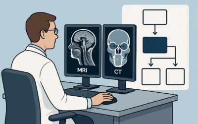
## Why Is MRI of the Head and Orbits the Recommended Initial Study?
The ACR designates both MRI head without and with IV contrast and MRI orbits without and with IV contrast as Usually Appropriate for the initial imaging of suspected optic neuritis. In practice, a combined or tailored protocol that includes high-resolution orbital sequences and full brain imaging is ideal. This approach provides the most comprehensive diagnostic information without exposing the patient to ionizing radiation (0 mSv).
The rationale for this recommendation is based on the superior soft-tissue contrast of MRI, which is essential for visualizing both the optic nerves and the brain parenchyma. The study is performed in two parts for distinct reasons:
- Without IV Contrast: Pre-contrast T2-weighted and Fluid-Attenuated Inversion Recovery (FLAIR) sequences of the brain are highly sensitive for detecting demyelinating plaques. The characteristic distribution of these lesions (e.g., periventricular, juxtacortical, infratentorial) is a cornerstone of the McDonald criteria for diagnosing MS.
- With IV Contrast: Post-contrast, fat-suppressed T1-weighted images are crucial for visualizing the optic nerves. Active inflammation causes a breakdown of the blood-nerve barrier, leading to gadolinium enhancement of the affected optic nerve segment. This finding confirms the diagnosis of optic neuritis. Contrast also helps identify any actively enhancing (i.e., new) demyelinating lesions in the brain.
An MRI orbits without IV contrast is also rated Usually Appropriate and can demonstrate optic nerve swelling on T2-weighted sequences, but it cannot confirm active inflammation and provides no information about brain lesions, limiting its utility.
Alternative studies are rated lower for clear reasons:
- CT of the head or orbits is rated Usually not appropriate. CT has poor sensitivity for both optic nerve inflammation and white matter plaques. It exposes the patient to unnecessary ionizing radiation (☢☢☢ 1-10 mSv) without providing the necessary diagnostic information for this clinical question.
- MRA or CTA of the head and neck is also rated Usually not appropriate. The primary concern in optic neuritis is inflammatory or demyelinating, not vascular. These studies are designed to evaluate for aneurysms, stenosis, or dissection and are not indicated for this scenario.
## What Are the Next Steps After an MRI for Suspected Optic Neuritis?
The results of the MRI will guide the subsequent clinical workflow, including treatment decisions, further testing, and specialist consultation.
- If the MRI is positive for optic nerve enhancement and shows brain lesions consistent with demyelination: This finding carries a high risk for the future development of MS. The patient should be started on high-dose corticosteroids (the standard treatment for acute optic neuritis to speed visual recovery) and referred to a neurologist for further evaluation. This may include a lumbar puncture for cerebrospinal fluid analysis and consideration of long-term disease-modifying therapy for MS.
- If the MRI shows optic nerve enhancement but the brain is normal: This confirms optic neuritis but is associated with a lower risk of developing MS. The patient is still treated with corticosteroids. However, if the clinical or imaging features are atypical (e.g., bilateral involvement, very long segment of enhancement), serologic testing for NMOSD (AQP4-IgG) and MOGAD (MOG-IgG) is warranted.
- If the MRI is entirely negative: A normal MRI makes typical demyelinating optic neuritis less likely, though not impossible, especially if imaging is performed very early or late in the clinical course. The clinician should reconsider the differential diagnosis, including ischemic, hereditary, or toxic/nutritional optic neuropathies. Further ophthalmologic evaluation and potentially a lumbar puncture may be necessary.
## Pitfalls to Avoid (and When to Get Help)
Navigating the workup for suspected optic neuritis requires careful attention to detail to avoid common diagnostic errors.
1. Ordering the wrong study: The most common pitfall is ordering a CT scan. CT lacks the sensitivity to answer the clinical question and results in a delayed diagnosis and unnecessary radiation exposure.
2. Omitting IV contrast: An MRI without gadolinium cannot confirm active inflammation in the optic nerve or brain, which is a critical piece of diagnostic and prognostic information. Always specify “without and with IV contrast.”
3. Not requesting orbital sequences: While a standard brain MRI is essential, dedicated fat-suppressed views of the orbits provide the best visualization of the optic nerves. Ensure your imaging order or protocol includes these sequences.
4. Ignoring atypical features: Red flags such as severe vision loss (no light perception), bilateral presentation, or lack of pain should prompt consideration of atypical optic neuritis (NMOSD, MOGAD) or alternative diagnoses and an expedited referral to neurology or neuro-ophthalmology.
If the clinical picture is complex or the imaging findings are ambiguous, consultation with a neurologist and/or a neuroradiologist is the appropriate next step.
## Related ACR Topics and Tools
For a comprehensive overview of all clinical variants related to orbital and visual pathway imaging, please see our parent guide. For tools to assist in ordering the correct study and understanding imaging protocols, the following resources are available.
- For breadth across all scenarios in Orbits, Vision, and Visual Loss, see our parent guide: Orbits, Vision, and Visual Loss: ACR Appropriateness Decoded.
- To explore adjacent clinical scenarios and their recommended imaging: ACR Appropriateness Criteria Lookup
- To review technical details for the recommended MRI study: Imaging Protocol Library
- For discussions about cumulative radiation exposure from prior studies: Radiation Dose Calculator
Frequently Asked Questions
Why is an MRI of the brain necessary if the symptoms are only in the eye?
While the symptoms are ocular, optic neuritis is frequently the first sign of a systemic neurologic disease, most commonly multiple sclerosis (MS). An MRI of the brain is the most sensitive tool for detecting the characteristic demyelinating lesions of MS. The presence of these lesions at the time of optic neuritis is the single most important factor in predicting the patient’s long-term risk of developing clinically definite MS.
Can I order an MRI of the orbits alone for suspected optic neuritis?
According to the ACR, an ‘MRI orbits without and with IV contrast’ is also rated ‘Usually Appropriate.’ It provides excellent high-resolution images of the optic nerves to confirm inflammation. However, it does not evaluate the brain for demyelinating plaques. Therefore, ordering an orbits-only study provides an incomplete picture for prognostication and long-term management. Most modern protocols combine dedicated orbital views with a full brain MRI.
Is gadolinium contrast always required for an optic neuritis workup?
Yes, intravenous gadolinium contrast is essential. While non-contrast T2-weighted images may show swelling and high signal in the optic nerve, post-contrast fat-suppressed T1-weighted images are needed to confirm active inflammation, which appears as enhancement. This confirmation is critical for the diagnosis. Contrast also helps identify new, active lesions in the brain.
What if my patient has a contraindication to MRI, like a non-compatible pacemaker?
This is a challenging situation, as CT is a poor substitute. In cases with an absolute contraindication to MRI, the diagnosis must rely more heavily on clinical findings, visual evoked potentials, and cerebrospinal fluid analysis from a lumbar puncture. A CT scan is rated ‘Usually not appropriate’ and is unlikely to be helpful. Consultation with neurology and ophthalmology is critical in these cases.
How soon after symptom onset should the MRI be performed?
The MRI should be performed as soon as is practical. Optic nerve enhancement is typically most prominent within the first two weeks of symptom onset and may begin to fade after 30 days. Imaging performed too late may miss the characteristic enhancement, potentially confusing the diagnosis. Prompt imaging ensures the highest diagnostic yield.
Reviewed by Pouyan Golshani, MD, Interventional Radiologist — May 29, 2026