Which Supplemental Screening Is Best for Intermediate-Risk Women with Dense Breasts?

A 48-year-old woman with a personal history of atypical ductal hyperplasia (ADH) presents for her annual wellness visit. Her lifetime risk of breast cancer is calculated at 18% via the Tyrer-Cuzick model, placing her in the intermediate-risk category. Her most recent mammogram report noted heterogeneously dense breasts (BI-RADS C). She has no new lumps or symptoms but is anxious about the masking effect of her breast density and asks what additional screening she should undergo. This common clinical question requires navigating the options for supplemental screening beyond standard mammography. According to the American College of Radiology (ACR) Appropriateness Criteria, for this specific scenario, Digital Breast Tomosynthesis (DBT) screening is rated Usually Appropriate.
Who Fits This Clinical Scenario?
This guidance applies to adult women who meet a specific combination of risk and breast density criteria. The key inclusion factors are:
- Intermediate Risk: This category includes women with a calculated lifetime risk of 15% to 20% for breast cancer. Common reasons for this risk level include a personal history of lobular carcinoma in situ (LCIS), atypical ductal hyperplasia (ADH), or atypical lobular hyperplasia (ALH). It can also be determined by risk assessment models (e.g., Tyrer-Cuzick) that incorporate family history and other factors.
- Heterogeneously Dense Breasts: This corresponds to Breast Imaging Reporting and Data System (BI-RADS) density category C. In these breasts, there are scattered areas of fibroglandular density which could obscure small masses on a standard 2D mammogram.
- Screening Context: The patient is asymptomatic, and the goal is supplemental screening to improve cancer detection, not to evaluate a clinical problem like a palpable lump or nipple discharge.
This workflow is distinct from other scenarios. It does not apply to women at average risk (<15% lifetime risk) or high risk (>20% lifetime risk, a known pathogenic genetic mutation, or a history of chest radiation therapy at a young age). Similarly, patients with nondense (BI-RADS A or B) or extremely dense (BI-RADS D) breasts fall under different recommendations. Applying this guidance to those groups would be inappropriate.
What Diagnoses Are You Working Up in This Scenario?
In supplemental screening for an asymptomatic, intermediate-risk woman with dense breasts, the primary goal is to detect occult malignancies that may be masked by the fibroglandular tissue on standard mammography. The “differential” is focused on early-stage, non-palpable disease.
Occult Invasive Ductal Carcinoma (IDC): This is the most common type of invasive breast cancer. In dense breasts, a small, developing IDC can be indistinguishable from normal glandular tissue on a 2D mammogram. Supplemental screening aims to identify these early cancers, often appearing as spiculated masses or areas of architectural distortion, before they become clinically apparent.
Occult Ductal Carcinoma In Situ (DCIS): DCIS is a non-invasive, stage 0 breast cancer. While often detected by calcifications on mammography, some forms of DCIS, particularly non-calcified or low-grade types, can be subtle. More sensitive screening modalities can improve the detection of DCIS that might otherwise be missed until it progresses.
High-Risk Precursor Lesions: While not frank malignancies, lesions like atypical ductal hyperplasia (ADH) or lobular carcinoma in situ (LCIS) confer an increased risk of future cancer. The patient in this scenario may already have a history of such a lesion, but supplemental screening can sometimes identify new, unsuspected areas that warrant biopsy and closer surveillance.
Why Is Digital Breast Tomosynthesis Screening the Recommended Study?
For an intermediate-risk woman with heterogeneously dense breasts, the ACR designates Digital Breast Tomosynthesis (DBT) screening as Usually Appropriate. DBT, often called 3D mammography, acquires multiple low-dose images of the breast from different angles, which are then reconstructed into thin, one-millimeter slices. This technique significantly mitigates the tissue-overlap problem inherent in 2D mammography, which is the primary cause of masking in dense breasts.
DBT improves the radiologist’s ability to distinguish a true lesion from superimposed normal tissue. This leads to a well-documented increase in cancer detection rates, particularly for invasive cancers, and a simultaneous reduction in the recall rate for false positives. The radiation dose for a combined 2D/3D mammogram (adult RRL ☢☢, 0.1-1 mSv) is slightly higher than 2D alone but remains well within accepted safety limits and is considered a favorable trade-off for the diagnostic benefits.
Other modalities are also considered:
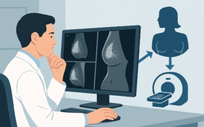
- Breast MRI with contrast (abbreviated or full protocol) is also rated Usually Appropriate. It is the most sensitive modality for detecting invasive cancer. However, it is more costly, requires IV contrast, and has a higher rate of benign biopsies compared to mammography. Its use in the intermediate-risk group is often a shared decision based on the specific degree of risk, patient preference, and insurance coverage.
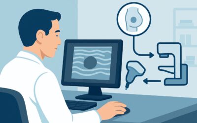
- Whole Breast Ultrasound (US) is rated May be appropriate. While it can detect additional cancers missed by mammography and uses no ionizing radiation, it is highly operator-dependent and has a significantly higher false-positive rate, leading to more unnecessary biopsies than DBT or MRI.
- MRI without IV contrast is rated Usually not appropriate. The administration of gadolinium-based contrast is essential for characterizing breast lesions and identifying cancers, which typically demonstrate enhancement. A non-contrast study has unacceptably low sensitivity for malignancy.
When ordering, specifying “Screening Mammogram with Tomosynthesis” is crucial. For detailed technical specifications, refer to our guide on the Screening Mammography (with DBT) protocol.
What’s Next After Digital Breast Tomosynthesis Screening? Downstream Workflow
The results of the DBT will guide the subsequent clinical pathway. The workflow branches based on the BI-RADS assessment provided in the radiology report.
- Negative or Benign Finding (BI-RADS 1 or 2): If the study is negative, the patient should continue with her routine annual screening schedule. No immediate further action is needed. The conversation about supplemental screening can be revisited at her next annual exam.
- Incomplete Finding (BI-RADS 0): This indicates the radiologist needs additional imaging to make a final assessment. The patient will be called back for diagnostic imaging, which typically includes diagnostic mammographic views (e.g., spot compression, magnification) and/or a targeted breast ultrasound of the area in question. This is a common outcome and should be explained to the patient as a necessary step for clarification, not a definitive sign of cancer.
- Suspicious Finding (BI-RADS 4 or 5): If the DBT identifies a lesion with suspicious features (e.g., a spiculated mass, suspicious calcifications, or significant architectural distortion), the report will recommend a biopsy. The next step is typically a referral for an image-guided core needle biopsy (stereotactic, tomosynthesis-guided, or ultrasound-guided, depending on the lesion’s characteristics) to obtain a tissue diagnosis.
- Probably Benign Finding (BI-RADS 3): This category is for findings that have a very high likelihood of being benign (>98%), but not definitively so. The standard recommendation is a short-interval follow-up, typically a diagnostic mammogram in six months, to ensure stability.
Pitfalls to Avoid (and When to Get Help)
Navigating supplemental screening requires careful attention to detail to avoid common missteps.
- Misclassifying Patient Risk: Incorrectly categorizing a high-risk patient (e.g., a BRCA gene carrier) as intermediate risk could lead to under-screening. Always use a formal risk model or clear high-risk criteria to ensure the patient is in the correct screening pathway.
- Ordering a “Screening” Study for a Symptomatic Patient: If the patient reports a new lump, skin change, or nipple discharge, the correct order is a diagnostic mammogram and ultrasound, not a screening study. Screening is strictly for asymptomatic individuals.
- Not Specifying Tomosynthesis (DBT): In some centers, DBT is not yet the default. Failing to explicitly request “with tomosynthesis” may result in a 2D-only mammogram, negating the primary benefit for a woman with dense breasts.
- Ignoring Insurance Preauthorization: While many states mandate coverage for supplemental screening for dense breasts, the specifics vary. Failing to confirm coverage or obtain preauthorization for modalities like MRI can result in unexpected costs for the patient.
If a biopsy is recommended (BI-RADS 4 or 5), immediate escalation to a breast surgeon or a dedicated breast imaging center for consultation and tissue sampling is the appropriate next step.
Related ACR Topics and Tools
This article covers one specific variant within the broader topic of supplemental breast cancer screening. For a comprehensive overview of all clinical scenarios, including different risk levels and breast densities, please consult the parent topic guide. The following GigHz tools can also support your clinical workflow:
- For breadth across all scenarios in Supplemental Breast Cancer Screening Based on Breast Density, see our parent guide: Supplemental Breast Cancer Screening Based on Breast Density: ACR Appropriateness Decoded.
- ACR Appropriateness Criteria Lookup — for quickly checking adjacent scenarios or different clinical questions.
- Imaging Protocol Library — for detailed technical guidance on performing the recommended studies.
- Radiation Dose Calculator — for discussing cumulative radiation exposure with patients.
Frequently Asked Questions
Why is Breast MRI also ‘Usually Appropriate’ for this scenario, and how do I choose between it and DBT?
Breast MRI with contrast is the most sensitive test for detecting invasive breast cancer and is also rated ‘Usually Appropriate.’ The choice between DBT and MRI for an intermediate-risk woman is a clinical judgment based on shared decision-making. Factors to consider include the patient’s specific lifetime risk (a woman at 19% risk may be a stronger candidate for MRI than one at 15%), patient anxiety, claustrophobia, contraindications to MRI or contrast, cost, and insurance coverage. DBT is often the more practical and accessible first choice for supplemental screening in this group.
What if my patient has heterogeneously dense breasts but is at average risk?
If the patient is at average risk (<15% lifetime risk) with heterogeneously dense breasts, the ACR recommendations change. In that scenario, supplemental screening with modalities like ultrasound or MRI is generally not recommended, and DBT remains the standard screening examination. This highlights the importance of accurate risk assessment, as risk level is a key determinant of the appropriateness of supplemental imaging.
Does a personal history of breast cancer change this recommendation?
Yes, significantly. A personal history of breast cancer moves the patient into a different clinical category: surveillance imaging, not screening. The ACR has separate guidelines for surveillance of women with a personal history of treated breast cancer, which typically involve annual mammography and often include annual supplemental breast MRI, regardless of breast density.
Is contrast-enhanced mammography (CEM) an option for this patient?
Contrast-enhanced mammography is rated ‘May be appropriate’ for this scenario. CEM uses iodinated IV contrast to highlight areas of abnormal blood flow, similar to MRI, but is performed on a mammography machine. It has shown sensitivity comparable to MRI in some studies and can be an alternative when MRI is contraindicated or unavailable. However, it is less widely available than DBT or MRI and involves both radiation and IV contrast.
How do I calculate a patient’s lifetime breast cancer risk to determine if she is intermediate risk?
Several validated statistical models are available to estimate lifetime breast cancer risk. The most commonly used in clinical practice is the Tyrer-Cuzick (IBIS) model, which incorporates family history in first- and second-degree relatives, personal reproductive history, history of prior benign breast biopsies, and other factors. Other models include the Gail model and the Claus model. Many EMRs and online calculators can facilitate this assessment.
Reviewed by Pouyan Golshani, MD, Interventional Radiologist — May 30, 2026