Your Patient Has a Traumatic Hip Fracture on Radiographs: What Imaging Should You Order Next?
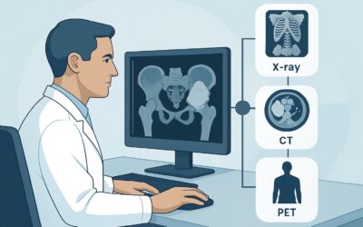
An 82-year-old woman is brought to the emergency department after a fall at home. She has severe right hip pain and cannot bear weight. Initial radiographs are performed, and the on-call radiologist confirms a displaced femoral neck fracture. The orthopedic surgeon is consulted and asks for more detailed imaging to plan for surgery. You are now faced with a critical decision: which advanced imaging study will provide the necessary anatomical detail to guide the operative plan, balancing speed, accuracy, and patient safety? This article provides a focused workflow for this exact scenario. According to the American College of Radiology (ACR) Appropriateness Criteria, CT hip without IV contrast is rated Usually Appropriate as the next imaging study.
Who Fits This Clinical Scenario for Post-Radiograph Hip Fracture Imaging?
This guidance is specifically for an adult patient who presents with acute hip pain following a clear traumatic event, and for whom initial radiographs have already confirmed the presence of a hip fracture. The primary clinical question is no longer if a fracture exists, but rather to characterize it fully for preoperative planning.
This workflow does not apply to several similar, but distinct, clinical situations:
- Suspected fracture with negative radiographs: If a patient has a high clinical suspicion for a hip fracture (e.g., inability to bear weight after a fall) but the initial X-rays are negative or indeterminate, the imaging pathway is different. That scenario requires a study with higher sensitivity for detecting occult fractures.
- Post-reduction of a hip dislocation: Imaging performed after the successful reduction of a hip dislocation has a different goal, primarily to assess for joint congruity, entrapped fragments, and associated fractures.
- Suspected soft tissue injury: If the primary clinical concern after trauma is a tendon, muscle, or ligament injury and radiographs are negative for fracture, the imaging workup would focus on soft tissue evaluation, typically with different modalities.
- Initial imaging: This article assumes radiographs have already been performed. For guidance on the very first imaging study to order for traumatic hip pain, a different ACR variant applies.
Correctly identifying your patient’s scenario is crucial for ordering the most effective and appropriate next study.
What Pre-Surgical Details Are You Clarifying with Advanced Imaging?
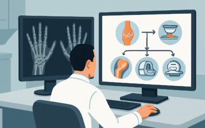
Once a hip fracture is identified on radiographs, the diagnosis is established. The purpose of subsequent imaging is not to re-diagnose but to refine the anatomical understanding for surgical planning. The key questions you are trying to answer with the next study directly influence the choice of operative approach and hardware.
Fracture Pattern and Comminution: Radiographs provide a two-dimensional view, which can underestimate the complexity of a fracture. Advanced imaging is needed to precisely delineate the fracture lines, determine the number and displacement of fragments (comminution), and assess for stability. This information is critical for the surgeon to decide between internal fixation (screws, plates, or an intramedullary nail) and arthroplasty (hemiarthroplasty or total hip replacement).
Articular Surface Involvement: A crucial detail is whether the fracture extends into the weight-bearing articular surface of the femoral head or the acetabulum. Intra-articular extension significantly impacts prognosis and demands meticulous anatomical reduction during surgery to minimize the risk of post-traumatic arthritis.
Occult Associated Fractures: Patients with a significant hip fracture, particularly from high-energy trauma, may have other, less obvious fractures in the pelvis (like pubic rami fractures) or proximal femur. Identifying these associated injuries is essential for a comprehensive surgical plan and to avoid missing injuries that could affect postoperative rehabilitation.
Pre-existing Bony Pathology: The advanced study can also reveal underlying conditions like severe osteoarthritis, avascular necrosis, or bone cysts that were not fully appreciated on the plain films. The presence of these conditions may lead the surgeon to choose a different procedure, such as a total hip arthroplasty instead of internal fixation, to address both the fracture and the underlying joint disease simultaneously.
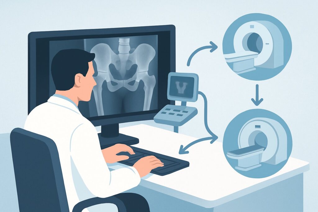
Why Is CT hip without IV contrast Usually Appropriate for Preoperative Planning?
The ACR designates CT hip without IV contrast as Usually Appropriate because it directly and efficiently answers the critical questions needed for surgical planning in a patient with a known hip fracture.
Computed Tomography (CT) provides exceptional high-resolution detail of cortical and trabecular bone, far surpassing that of radiographs. Its ability to generate multiplanar reformats and 3D reconstructions gives surgeons an unparalleled understanding of the fracture’s spatial relationships, displacement, and comminution. This level of detail is often the deciding factor in selecting the optimal surgical hardware and approach. Furthermore, CT is fast and widely available in most emergency settings, which is a key consideration for a patient in pain awaiting surgery.
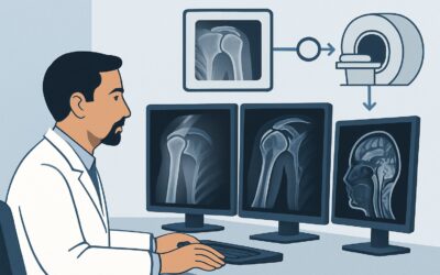
Why are alternative studies rated lower for this specific scenario?
- MRI hip without IV contrast: While rated as May be appropriate, MRI is generally not the first choice for preoperative planning of a known fracture. Its primary strength is in evaluating soft tissues and detecting occult fractures or bone marrow edema, which is not the main clinical question here. For pure bony characterization, it is slower, more expensive, less readily available, and more susceptible to motion artifact than CT. The superior bone detail of CT is the priority.
- CT hip with IV contrast: This study is rated Usually not appropriate. Intravenous contrast is used to evaluate vascular structures, soft tissue masses, and infection. It adds no value to the assessment of bony fracture patterns. Ordering it unnecessarily exposes the patient to the risks of contrast media (allergic reaction, contrast-induced nephropathy) without providing any additional benefit for this indication.
The recommended study, CT hip without IV contrast, carries a moderate radiation dose (ACR Relative Radiation Level ☢☢☢, corresponding to 1-10 mSv). However, in the context of an adult with a major traumatic injury requiring surgery, the clinical benefit of obtaining precise anatomical information to ensure a successful operation far outweighs the radiation risk.
What Happens After the CT Scan? Interpreting Results for Surgical Planning
The results of the preoperative CT scan directly guide the orthopedic surgeon’s next steps. The detailed report and images will trigger a specific surgical pathway.
- If the CT confirms a displaced femoral neck fracture in an older adult: The detailed view of bone quality and fracture pattern helps the surgeon decide between hemiarthroplasty (replacing the femoral head) and total hip arthroplasty (replacing both the head and the socket). The CT data can also be used for preoperative digital templating to select the appropriate implant size.
- If the CT shows a complex, comminuted intertrochanteric or subtrochanteric fracture: The 3D reconstructions are invaluable for planning the placement of an intramedullary nail or a plate-and-screw construct. The surgeon can anticipate the need for specific reduction techniques or bone grafting based on the fracture’s complexity.
- If the CT reveals significant acetabular involvement: This finding changes the entire scope of the operation. An isolated hip fracture is managed differently than a combined hip and acetabular fracture. This result may prompt a consultation with a trauma surgeon specializing in pelvic and acetabular reconstruction and will necessitate a more complex surgical approach (e.g., open reduction and internal fixation).
- If the CT identifies unexpected findings: The discovery of pre-existing severe arthritis or avascular necrosis may shift the plan from fracture fixation to a total hip replacement to address both problems in a single surgery, improving the patient’s long-term outcome.
Pitfalls to Avoid (and When to Get Help)
Even in this relatively straightforward scenario, several pitfalls can compromise patient care.
- Ordering with IV Contrast: A common error is ordering a “CT hip” without specifying “without IV contrast.” This unnecessarily adds risk and cost. Always be explicit in the order.
- Delaying the Scan: For displaced fractures, particularly of the femoral neck, timely surgery is associated with better outcomes. Unnecessary delays in obtaining the preoperative CT can postpone surgery, increasing complication rates.
- Ignoring the Rest of the Pelvis: Ensure the scan protocol includes the entire pelvis if there is any suspicion of associated injuries. A scan limited to just the hip might miss a concurrent sacral or pubic ramus fracture.
- Misinterpreting the Patient Scenario: Applying this workflow to a patient with a negative radiograph is incorrect. If the X-ray is negative but suspicion remains high, the appropriate next step is typically an MRI, not a CT.
If the CT reveals highly complex fracture patterns, such as extension into the sacroiliac joints or significant pelvic ring disruption, immediate escalation to an orthopedic trauma specialist with expertise in pelvic surgery is warranted.
Related ACR Topics and Tools
For a comprehensive overview of imaging for all acute hip pain scenarios, from trauma to atraumatic causes, please see our parent guide. For tools to assist in ordering and interpreting imaging, the following resources are available.
- Parent Topic Hub: For breadth across all scenarios in Acute Hip Pain, see our parent guide: Acute Hip Pain: ACR Appropriateness Decoded.
- ACR Criteria Lookup: To explore other clinical variants or confirm appropriateness for different patient presentations, use the ACR Appropriateness Criteria Lookup.
- Imaging Protocols: For detailed technical parameters of CT and other imaging studies, consult the Imaging Protocol Library.
- Radiation Dose: To discuss radiation exposure with patients, the Radiation Dose Calculator can help contextualize the dose from a CT scan.
Frequently Asked Questions
Why not just go straight to the operating room based on the X-ray?
For some simple, non-displaced fractures, surgery might be planned from radiographs alone. However, for most displaced or potentially complex fractures, X-rays do not provide sufficient detail. A preoperative CT scan helps the surgeon understand the three-dimensional fracture anatomy, identify comminution, and plan the precise placement of hardware, which can lead to shorter operative times, less blood loss, and better surgical outcomes.
Is MRI ever the right choice for preoperative planning of a known hip fracture?
While CT is ‘Usually Appropriate,’ the ACR rates MRI as ‘May be appropriate.’ MRI might be considered in rare, specific circumstances, such as a strong suspicion of associated soft-tissue pathology (like a gluteal tendon tear) that would alter the surgical approach, or in a younger patient where assessing the viability of the femoral head is a primary concern. However, for routine bony characterization, CT is faster and provides superior bone detail.
Does the patient’s age change the recommendation for CT?
This ACR variant is for adults, and the recommendation for CT is generally applicable across the adult age spectrum. While radiation exposure is always a consideration, the immediate benefit of accurate surgical planning for a major, life-altering injury like a hip fracture is considered to outweigh the long-term stochastic risk of radiation in an adult patient.
What if the patient has renal insufficiency? Is a non-contrast CT still safe?
Yes. A CT scan performed without intravenous contrast media poses no risk to kidney function. The recommendation for ‘CT hip without IV contrast’ is specifically chosen to avoid the risks associated with contrast agents, making it a safe choice for patients with pre-existing renal insufficiency.
If the radiograph shows a hip dislocation with a fracture, does this guidance still apply?
This scenario is more complex. A fracture-dislocation is a true orthopedic emergency. The first priority is urgent reduction of the dislocation. Imaging guidance would then follow the ‘Post reduction of hip dislocation’ ACR variant, which typically involves a post-reduction CT scan to assess for joint congruity, intra-articular bone fragments, and to fully characterize the associated fracture.
Reviewed by Pouyan Golshani, MD, Interventional Radiologist — May 30, 2026