What Is the Next Imaging Study for Chronic Shoulder Pain With Osteoarthritis on Radiographs?
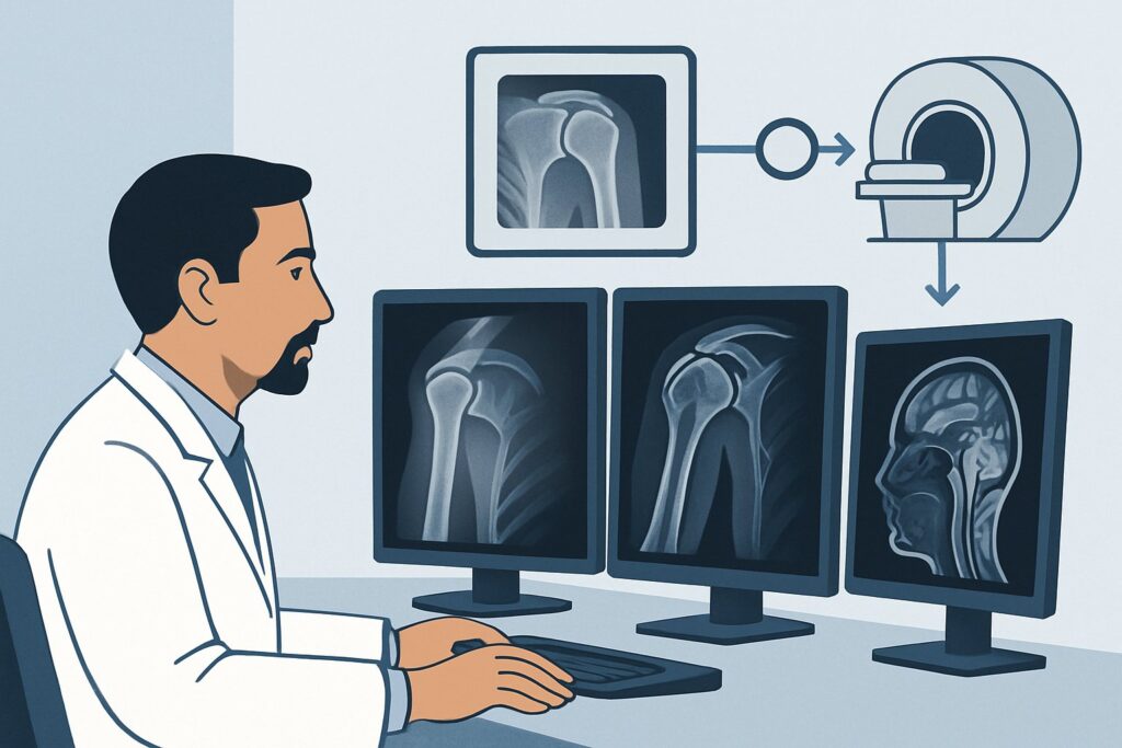
A 62-year-old patient with a history of manual labor presents with several months of worsening, deep right shoulder pain and mechanical symptoms. You’ve already obtained initial radiographs, which confirm moderate to severe glenohumeral osteoarthritis with osteophyte formation and joint space narrowing. The patient’s function is now significantly limited, and you need to determine the next steps in management, which may include surgery. What is the most appropriate next imaging study to order to fully assess the shoulder and guide treatment?
This clinical workflow article addresses this specific scenario. According to the American College of Radiology (ACR) Appropriateness Criteria, for a patient with chronic shoulder pain and known osteoarthritis on radiographs, an MRI shoulder without IV contrast is rated Usually Appropriate as the next imaging study. This guide details the rationale, differential diagnosis, and downstream decision-making for this common clinical presentation.
Who Fits This Clinical Scenario for Chronic Shoulder Pain?
This guidance is specifically for adult patients with chronic shoulder pain (typically lasting weeks to months) where the initial diagnostic step—plain radiographs—has already been completed and has demonstrated findings of osteoarthritis. The key element is that the bony diagnosis is established, and the clinical question has evolved to assessing the soft tissue structures to guide further treatment, particularly pre-operative planning.
This workflow applies if your primary goal is to evaluate the integrity of the rotator cuff, assess the degree of cartilage loss, and visualize other soft tissue structures in the context of known bony degenerative disease.
This article does not apply to several similar-but-distinct clinical situations:
- Initial Imaging: If the patient has not yet had any imaging, the workup starts with radiographs. This scenario is covered in the initial imaging variant of the ACR criteria.
- Acute Trauma: Patients with an acute shoulder injury or suspected fracture follow a different diagnostic algorithm, often starting with a dedicated trauma series of radiographs.
- Primary Suspicion of Instability: In a patient where the main concern is a labral tear or instability (e.g., a younger patient with recurrent dislocations) and radiographs are unrevealing, MR arthrography may be considered more strongly.
- Calcific Tendinopathy on Radiographs: If the initial radiographs showed calcific deposits in the rotator cuff tendons as the primary finding, the subsequent imaging choice and management pathway differ from that of primary osteoarthritis.
Correctly identifying your patient’s specific scenario is crucial for ordering the most effective and appropriate imaging study.
What Diagnoses Are You Working Up When Radiographs Show Osteoarthritis?
Once radiographs confirm glenohumeral osteoarthritis, the clinical focus shifts from diagnosing the bony condition to understanding its consequences and identifying co-existing pathologies that contribute to the patient’s pain and functional decline. The next imaging study is not to re-diagnose the OA, but to complete the picture for treatment planning.
Rotator Cuff Tear (Full or Partial Thickness)
This is the most common and clinically significant co-existing pathology. Degenerative rotator cuff tears are frequently associated with advanced osteoarthritis. The integrity of the rotator cuff is the single most important factor in determining the type of shoulder arthroplasty a patient may need. A functional cuff allows for an anatomic total shoulder replacement, while a massive, irreparable tear often necessitates a reverse total shoulder arthroplasty.
Rotator Cuff Muscle Atrophy and Fatty Infiltration
Beyond a simple tear, the chronic disuse and altered mechanics from a rotator cuff tear lead to muscle atrophy and fatty infiltration. Assessing the quality of the remaining muscle (e.g., using the Goutallier classification) is critical for predicting the functional outcome of a potential repair and influences surgical decision-making.
Biceps Tendon Pathology
The long head of the biceps tendon is intimately related to the rotator cuff and is often implicated in shoulder pain. The next imaging study aims to identify tendinosis, partial tears, or subluxation/dislocation of the tendon from the bicipital groove, all of which are common in the setting of degenerative shoulder disease.
Detailed Cartilage and Bone Assessment
While radiographs show joint space narrowing, they provide a limited two-dimensional view. Advanced imaging offers a detailed, multiplanar assessment of the location and severity of cartilage loss, the presence of subchondral cysts, and the precise morphology of osteophytes. It also allows for evaluation of glenoid bone stock and version, which are vital parameters for surgical planning.
Why Is MRI Shoulder Without Contrast the Recommended Next Study?
For a patient with known osteoarthritis on radiographs, the ACR panel rates MRI shoulder without IV contrast as Usually Appropriate. This recommendation is based on its superior ability to answer the key clinical questions that arise after the initial bony diagnosis is made.
The primary rationale is MRI’s excellent soft-tissue contrast. It provides a detailed, non-invasive evaluation of all the critical structures that will dictate treatment: the rotator cuff tendons, the associated muscles, the biceps tendon, the labrum, and the articular cartilage. It can reliably distinguish partial-thickness from full-thickness rotator cuff tears and quantify muscle atrophy and fatty infiltration, information that is essential for pre-operative planning and cannot be obtained from radiographs.
Let’s examine why other modalities are rated lower for this specific scenario:
- US shoulder is rated Usually not appropriate. While ultrasound is a valuable tool for assessing the rotator cuff in many other contexts, its utility is significantly diminished in patients with advanced osteoarthritis. The large osteophytes common in OA can create acoustic shadowing, which physically blocks the ultrasound beam from visualizing the underlying tendons, leading to a non-diagnostic or incomplete examination.
- CT shoulder without IV contrast is rated May be appropriate. CT provides outstanding detail of the bone, surpassing radiographs in its ability to assess glenoid version, bone loss, and complex osteophyte morphology. It is a valuable tool for pre-operative planning, especially if there is concern for severe glenoid bone deficiency. However, its poor soft-tissue contrast makes it inferior to MRI for evaluating the rotator cuff and other soft tissues. CT is often reserved for patients with contraindications to MRI (e.g., an incompatible implanted device) or as a complementary study focused on bone stock assessment.
From a safety perspective, MRI without contrast avoids both ionizing radiation (RRL=O 0 mSv) and the potential risks associated with gadolinium-based contrast agents. An MRI shoulder without and with IV contrast is rated Usually not appropriate because, in the workup of degenerative OA, intravenous contrast typically adds little to no diagnostic value for assessing the key structures of interest (rotator cuff, muscle quality).
Once you’ve decided on MRI shoulder without contrast, our protocol guide covers the technique, contrast, and reading principles: MRI Shoulder Without Contrast.
What’s Next After the MRI? Downstream Workflow
The results of the shoulder MRI will directly guide the next steps in management, branching into non-operative and operative pathways. The report provides the comprehensive structural information needed to have an informed discussion with the patient and, if necessary, a surgical consultant.
If the MRI confirms a large, full-thickness rotator cuff tear with significant muscle atrophy:
This finding often pushes the treatment algorithm toward surgical intervention, specifically a reverse total shoulder arthroplasty, as a standard anatomic replacement is likely to fail. The patient should be referred to an orthopedic surgeon to discuss the risks and benefits of this procedure.
If the MRI shows intact or only mildly degenerated rotator cuff tendons:
This is a more favorable prognostic sign. The patient may be a candidate for an anatomic total shoulder arthroplasty if their pain and functional limitation are severe enough to warrant surgery. Alternatively, they may continue with conservative management, including physical therapy, activity modification, and intra-articular injections, with the knowledge that the primary structural support of the shoulder is intact.
If the MRI is negative for a significant rotator cuff tear but shows other pathology (e.g., severe biceps tendinopathy, a degenerative labral tear):
These findings can be addressed with targeted treatments. The patient might benefit from an image-guided corticosteroid injection (rated May be appropriate by the ACR) into the biceps tendon sheath or subacromial bursa to see if it provides relief. If conservative measures fail, arthroscopic procedures like a biceps tenotomy or tenodesis could be considered.
If the MRI findings are discordant with the clinical picture:
In rare cases, the degree of pathology on MRI may not seem to explain the severity of the patient’s symptoms. In this situation, a diagnostic intra-articular anesthetic injection can be a useful tool to confirm that the glenohumeral joint is indeed the primary pain generator before proceeding with a major surgery like arthroplasty.
Pitfalls to Avoid (and When to Get Help)
Navigating the workup for chronic shoulder pain requires careful attention to the specific clinical context. Here are a few common pitfalls to avoid in this scenario:
- Ordering Ultrasound First: After radiographs show significant OA, proceeding directly to ultrasound is a common misstep. The likely presence of osteophytes makes ultrasound a low-yield study in this setting, potentially delaying the definitive diagnosis that an MRI would provide.
- Skipping Radiographs: Never order an MRI for chronic, non-traumatic shoulder pain without first obtaining plain radiographs. Radiographs are inexpensive, fast, and provide the essential bony context needed to interpret advanced imaging and select the right study.
- Using Contrast Unnecessarily: For a standard degenerative OA workup, ordering an MRI “with and without contrast” is generally not indicated. This adds cost, time, and the small risk of a contrast reaction without providing additional information relevant to the primary clinical questions.
- Ignoring MRI Contraindications: Always screen patients for contraindications to MRI, such as certain pacemakers, cochlear implants, or other metallic foreign bodies. If a patient cannot safely undergo an MRI, CT is the appropriate alternative for advanced bony assessment.
If the imaging findings are complex or the optimal surgical approach is unclear, an early consultation with an orthopedic surgeon specializing in shoulder surgery is the most appropriate next step.
Related ACR Topics and Tools
For a comprehensive overview of imaging for all chronic shoulder pain presentations, and for tools to help you choose the right study, the following resources are available:
- Parent Topic Hub Article: For breadth across all scenarios in Chronic Shoulder Pain, see our parent guide: Chronic Shoulder Pain: ACR Appropriateness Decoded.
- ACR Criteria Lookup: To explore adjacent scenarios or other clinical topics, use the ACR Appropriateness Criteria Lookup.
- Imaging Protocol Library: For technical details on how specific studies are performed, visit the Imaging Protocol Library.
- Radiation Dose Calculator: To discuss cumulative radiation exposure with patients for studies that use ionizing radiation, see the Radiation Dose Calculator.
Frequently Asked Questions
Why not order a CT scan instead of an MRI if I’m planning for surgery?
A CT scan is excellent for evaluating bone stock, glenoid version, and complex bony anatomy, and it is often used by surgeons for pre-operative planning. However, it provides very limited information about the rotator cuff and other soft tissues. The ACR rates MRI as ‘Usually Appropriate’ because it answers the critical soft tissue questions (especially rotator cuff integrity) that determine the *type* of surgery needed (anatomic vs. reverse arthroplasty). A CT may be ordered in addition to an MRI or as an alternative if MRI is contraindicated.
Is an MR arthrogram better than a non-contrast MRI in this scenario?
No. The ACR rates MR arthrography as ‘May be appropriate’ but non-contrast MRI as ‘Usually Appropriate’. An MR arthrogram, which involves injecting contrast directly into the joint, is superior for evaluating subtle labral tears or capsular structures, which are the primary concern in younger patients with instability. In the context of advanced osteoarthritis, the main questions revolve around the rotator cuff and muscle quality, which are well-visualized on a standard non-contrast MRI.
My patient has a pacemaker. What is the best alternative to an MRI?
If a patient has a non-MRI-conditional pacemaker or another absolute contraindication to MRI, a CT shoulder without IV contrast is the best alternative for advanced imaging. While it won’t show the rotator cuff tendons well, it provides excellent bony detail for surgical planning. In some cases, a high-quality ultrasound performed by an experienced sonographer may provide some information about the rotator cuff, but this can be limited by bony osteophytes.
What if the MRI shows osteoarthritis but the rotator cuff is completely normal?
This is a positive finding. It indicates that the patient’s pain is likely driven by the glenohumeral arthritis itself, without the confounding factor of a rotator cuff tear. If conservative management fails, this patient would be an excellent candidate for a standard anatomic total shoulder arthroplasty, which generally has better functional outcomes and fewer complications than a reverse arthroplasty.
Should I order additional radiographic views before getting an MRI?
Generally, no. The ACR rates ‘Radiography shoulder additional views’ as ‘Usually not appropriate’ in this scenario. A standard set of initial radiographs (like an AP and an axillary lateral view) is usually sufficient to diagnose osteoarthritis. Once OA is confirmed, additional plain films provide diminishing returns. The next logical step for a comprehensive assessment is cross-sectional imaging with MRI.
Reviewed by Pouyan Golshani, MD, Interventional Radiologist — May 30, 2026