What Imaging Should You Order for a Suspected Mesenteric or Renal Embolic Source?

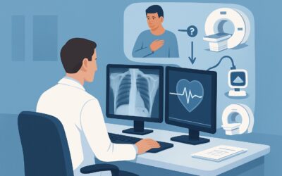
A 48-year-old patient presents to the emergency department with sudden, severe left flank pain. A contrast-enhanced computed tomography (CT) scan of the abdomen and pelvis reveals a classic wedge-shaped perfusion defect in the left kidney, diagnostic of a renal infarct. The patient has no history of trauma, vasculitis, or known hypercoagulable state, raising strong suspicion for an embolic etiology. The clinical team now faces a critical decision: what is the most effective next imaging study to identify the embolic source and guide treatment? This article provides a detailed workflow for this specific scenario, grounded in the American College of Radiology (ACR) Appropriateness Criteria, which rate `US echocardiography transesophageal` as `Usually appropriate` for this workup.
Who Fits This Clinical Scenario?
This guidance applies specifically to patients with an objectively confirmed arterial occlusion or infarct within the mesenteric or renal arterial systems, where the clinical suspicion for an embolic source is high. This includes presentations such as:
- Acute renal infarct discovered on CT or MRI
- Acute mesenteric ischemia attributed to a distal arterial embolus (not thrombosis of the main superior mesenteric artery)
- Splenic infarcts of suspected embolic origin
The key element is the need to identify the source of a presumed embolus that has already caused visceral organ damage. This workflow is distinct from and should not be applied to other related, but different, clinical situations. For example, this guidance does not cover:
- Isolated peripheral limb ischemia: A patient presenting with an acutely ischemic upper or lower extremity has a different diagnostic algorithm, covered in a separate ACR variant.
- Multiorgan system occlusions: When emboli are found in multiple, disparate vascular beds (e.g., renal, splenic, and cerebral), the workup may need to be broader, potentially prioritizing evaluation of the entire thoracic aorta.
- Known embolic source: In a patient with known atrial fibrillation and subtherapeutic anticoagulation, the source is often presumed, and imaging may be focused on characterizing the extent of end-organ damage rather than finding the source.
What Diagnoses Are You Working Up in This Scenario?
When a renal or mesenteric embolus is identified, the imaging workup is a search for the “smoking gun”—the primary source from which the clot or debris originated. The differential diagnosis for the embolic source is centered on cardiac and aortic pathologies.
Cardioembolic Source: This is the most common origin of systemic arterial emboli. The primary culprits are thrombi forming within the heart chambers or on the valves. This includes left atrial appendage thrombus, a frequent complication of atrial fibrillation (even if paroxysmal and not yet diagnosed). Another key consideration is a left ventricular thrombus, which can form over an area of akinesis or aneurysm, often following a myocardial infarction. Valvular pathologies, particularly infectious endocarditis with mobile vegetations, are also a high-priority source to identify or exclude.
Aortic Atheroembolism: Complex, ulcerated, or mobile atherosclerotic plaque within the thoracic or abdominal aorta can be a significant source of emboli. Fragments of plaque or overlying thrombus can break off (a “shaggy aorta”) and travel downstream to the visceral arteries. This is a particularly important consideration in older patients with extensive vascular risk factors.
Paradoxical Embolism: Though less common, this diagnosis is critical to consider, especially in younger patients without traditional risk factors for arterial disease. In this scenario, a thrombus from the venous system (like a deep vein thrombosis) travels to the right side of the heart and crosses to the left side through a patent foramen ovale (PFO) or an atrial septal defect (ASD), subsequently entering the systemic arterial circulation.
Why Is Transesophageal Echocardiography Usually Appropriate for a Suspected Embolic Source?
For a patient with a confirmed renal or mesenteric infarct, the ACR designates several imaging modalities as `Usually appropriate`, but transesophageal echocardiography (TEE) offers distinct advantages in visualizing the most likely high-yield sources. The rationale for its high rating is rooted in its superior diagnostic capability for key cardiac structures that are often poorly visualized by other non-invasive means.
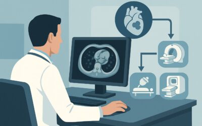
TEE involves passing an ultrasound probe into the esophagus, which provides an unobstructed acoustic window to the posterior structures of the heart. This is critical for evaluating the left atrial appendage—the most common site of thrombus formation in atrial fibrillation—which is often difficult to see clearly on a standard transthoracic echocardiogram (TTE). TEE also provides high-resolution images of the mitral and aortic valves to detect small vegetations of endocarditis, and it can clearly visualize the interatrial septum to assess for a PFO with a bubble study.
While `US echocardiography transthoracic resting` (TTE) is also rated `Usually appropriate` and is often the initial test due to its non-invasive nature, it may not be sufficient. TTE is excellent for assessing overall left ventricular function and identifying large, apical LV thrombi, but its sensitivity for left atrial appendage thrombus and small valvular vegetations is lower than TEE. Therefore, a negative TTE does not fully exclude a cardioembolic source, and TEE is often required for a definitive workup.
Other `Usually appropriate` studies include `CTA chest and abdomen with IV contrast` (Adult RRL=☢☢☢☢ 10-30 mSv) and `MRA chest and abdomen without and with IV contrast` (Adult RRL=O 0 mSv). These are excellent for evaluating the aorta for complex atheromatous plaque, aneurysm, or dissection as an embolic source. However, they provide limited functional and morphological detail of the heart itself. The choice between TEE and a CTA/MRA often depends on the pre-test probability of a cardiac versus an aortic source. In many cases, both cardiac and aortic imaging are ultimately required if the initial study is negative.
What’s Next After Echocardiography? Downstream Workflow for Renal or Mesenteric Embolism
The results of the initial imaging study will dictate the subsequent clinical pathway, focusing on treatment and secondary prevention.
- If TEE is positive for a definite source: A finding of a left atrial appendage thrombus, a mobile LV thrombus, or valvular vegetation solidifies the diagnosis. The immediate next step is to initiate appropriate therapy, which typically involves therapeutic anticoagulation to prevent further embolization. In cases of infectious endocarditis, urgent consultation with infectious disease and cardiothoracic surgery is warranted. The focus shifts from diagnosis to treatment and management of the underlying condition (e.g., rate/rhythm control for atrial fibrillation, post-MI care, or antibiotic/surgical management for endocarditis).
- If TEE is negative: A negative high-quality TEE makes a cardioembolic source less likely, though not impossible. The workup should then pivot to evaluating the aorta. The next logical step would be to order a `CTA chest and abdomen with IV contrast` or an `MRA chest and abdomen without and with IV contrast`, both rated `Usually appropriate`, to search for complex aortic plaque, mobile thrombus, or other aortic pathology. If this study is also negative, further investigation for a hypercoagulable state or rare causes like a cardiac tumor (myxoma) may be considered.
- If TEE is indeterminate or suggestive of a PFO: If a PFO is identified, especially in a younger patient with no other source, the diagnosis of paradoxical embolism becomes more probable. This may prompt a search for a deep vein thrombosis (DVT) with lower extremity duplex ultrasound and a discussion about PFO closure.
Pitfalls to Avoid When Investigating a Visceral Embolic Source
Navigating the workup for a suspected embolic source requires careful consideration to avoid common diagnostic traps.
- Stopping the workup after a negative TTE: A transthoracic echocardiogram is a good screening tool but is not sensitive enough to rule out key sources like a left atrial appendage thrombus. If clinical suspicion is high, a negative TTE should be followed by a TEE.
- Overlooking the aorta: While the heart is the most common source, the aorta is a frequent culprit, especially in patients with widespread atherosclerosis. Failing to image the thoracic and abdominal aorta after a negative cardiac workup is a significant pitfall.
- Ignoring patient contraindications: TEE is semi-invasive and may be contraindicated in patients with esophageal strictures, varices, or recent surgery. CTA requires iodinated contrast, which may be contraindicated in patients with severe renal insufficiency or allergy. MRA requires gadolinium-based contrast agents, which carry a risk of nephrogenic systemic fibrosis in patients with poor renal function. Always tailor the imaging choice to the individual patient’s clinical status and comorbidities.
If the initial workup is unrevealing despite a high clinical suspicion of an embolic event, escalation to a multidisciplinary team including cardiology, vascular surgery, and hematology is often the most prudent next step.
Related ACR Topics and Tools
For a comprehensive overview of all clinical variants related to this topic, please consult the parent guide. Additionally, several GigHz tools can assist in applying these criteria and communicating with patients about the imaging studies involved.
- For breadth across all scenarios in Workup of Noncerebral Systemic Arterial Embolic Source, see our parent guide: Workup of Noncerebral Systemic Arterial Embolic Source: ACR Appropriateness Decoded.
- To explore other clinical scenarios and their corresponding ACR ratings, use the ACR Appropriateness Criteria Lookup.
- For detailed procedural techniques on recommended studies, refer to the Imaging Protocol Library.
- To discuss radiation exposure with patients, especially when considering CT, the Radiation Dose Calculator can be a helpful resource.
Frequently Asked Questions
Why not start with a CTA of the chest and abdomen, since it’s also rated ‘Usually appropriate’?
Starting with a CTA is a reasonable alternative, especially if the patient’s risk profile strongly suggests an aortic source (e.g., elderly patient with severe, diffuse atherosclerosis). However, cardiac sources are statistically more common overall. TEE is more sensitive for the highest-yield cardiac findings like left atrial appendage thrombus and endocarditis vegetations. The choice often depends on clinical judgment, but a comprehensive workup frequently requires evaluation of both the heart and the aorta if the first study is negative.
What if my patient cannot tolerate a transesophageal echocardiogram (TEE)?
If a patient has contraindications to TEE (e.g., esophageal pathology) or refuses the procedure, the next best steps are other ‘Usually appropriate’ modalities. A cardiac MRI (CMR) can provide excellent morphological and functional data, including visualization of LV thrombus, without requiring sedation or an esophageal probe. A high-quality cardiac CT can also be very effective. These studies are excellent alternatives for assessing cardiac anatomy when TEE is not an option.
Is a transthoracic echocardiogram (TTE) ever sufficient for this workup?
A TTE may be sufficient if it reveals an unequivocal, high-risk embolic source, such as a large, mobile left ventricular thrombus in a patient with a recent myocardial infarction. In this case, finding the source on TTE obviates the need for a more invasive TEE. However, if the TTE is negative or equivocal, it is not sensitive enough to rule out a cardioembolic source, and further imaging with TEE is typically required.
Does finding a patent foramen ovale (PFO) on TEE confirm a paradoxical embolism?
Not necessarily. A PFO is a common incidental finding in up to 25% of the general population. To implicate it as the cause of a paradoxical embolus, you typically need to demonstrate a right-to-left shunt (often with a bubble study), have evidence of a venous thrombus (like a DVT), and have excluded other more common cardioembolic and aortic sources. The diagnosis is often one of inference after a thorough workup.
The initial CT that found the renal infarct used IV contrast. Is it safe to give more contrast for a CTA of the chest and abdomen soon after?
This is a critical clinical consideration. The risk of contrast-induced nephropathy (CIN) depends on the patient’s baseline renal function, the time between studies, and the total contrast volume. In a patient with normal renal function, the risk is low. In a patient with pre-existing chronic kidney disease, the risk is higher. It is essential to assess the patient’s eGFR, ensure adequate hydration, and weigh the diagnostic urgency of the CTA against the risk of further kidney injury. An MRA with gadolinium or a non-contrast study like TEE might be safer alternatives in high-risk patients.
Reviewed by Pouyan Golshani, MD, Interventional Radiologist — May 30, 2026