


Which Imaging Study Is Best for a Child with Neck Pain After Trauma?
ACR-guided imaging workflow for Child, 3 to 16 years of age, acute cervical spine trauma, at least one risk factor with reliable clinical examination (base
Part of the imaging decision system doctors were never taught to navigate at speed: which study to order, which protocol, and what the published appropriateness guidance actually says for pediatric imaging. Plain-English references, scenario by scenario.
ACR-guided imaging workflow for Child, 3 to 16 years of age, acute cervical spine trauma, at least one risk factor with reliable clinical examination (base

ACR-guided imaging workflow for Child, 3 to 16 years of age, acute cervical spine trauma, meets low risk criteria (based on PECARN or NEXUS). Initial imagi

ACR-guided imaging workflow for Child. Suspected acute appendicitis with clinical suspicion or initial imaging suggestive of complication (eg, abscess, bow
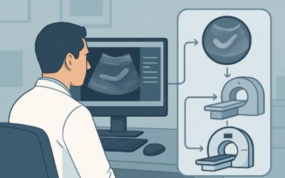
ACR-guided imaging workflow for Child. Suspected acute appendicitis, equivocal or nondiagnostic right lower quadrant ultrasound. Next imaging study. (Suspe

ACR-guided imaging workflow for Child. Suspected acute appendicitis, high clinical risk. Initial imaging. (Suspected Appendicitis-Child): recommended study

ACR-guided imaging workflow for Child. Suspected acute appendicitis, intermediate clinical risk. Initial imaging. (Suspected Appendicitis-Child): recommend

ACR-guided imaging workflow for Child. Suspected acute appendicitis, low clinical risk. Initial imaging. (Suspected Appendicitis-Child): recommended study,

ACR-guided imaging workflow for Adolescent (10 to 17 years of age). Adolescent idiopathic scoliosis. Risk factors. Initial imaging. (Scoliosis-Child): reco

ACR-guided imaging workflow for Adolescent (10 to 17 years of age). Adolescent idiopathic scoliosis. No risk factors. Initial imaging. (Scoliosis-Child): r

ACR-guided imaging workflow for Child (0 to 9 years of age). Early onset idiopathic scoliosis. Initial imaging. (Scoliosis-Child): recommended study, diffe