How Should You Work Up an Indeterminate Mediastinal Mass Found on PET/CT?

A 62-year-old patient with a history of non-small cell lung cancer, treated two years ago, undergoes routine surveillance imaging. The Fluorodeoxyglucose (FDG) Positron Emission Tomography/Computed Tomography (PET/CT) reveals a new, 3 cm prevascular mediastinal mass that is moderately FDG-avid. While concerning for recurrence, the morphology is atypical, and inflammatory causes cannot be excluded. The multidisciplinary tumor board is asking for the next step to secure a definitive diagnosis before committing to further therapy. This article details the American College of Radiology (ACR) guided workflow for choosing the next study for an indeterminate mediastinal mass on FDG-PET/CT. For this scenario, the ACR rates ‘Image-guided transthoracic needle biopsy’ as ‘Usually Appropriate’.
Who Fits This Clinical Scenario?
This guidance applies to a specific and common clinical decision point: a patient who has already undergone an FDG-PET/CT that has identified a mediastinal mass, but the findings remain equivocal. The key inclusion criterion is that the combination of anatomic features on CT and metabolic activity on PET is insufficient to make a confident diagnosis. This often occurs when a mass is FDG-avid, raising suspicion for malignancy, but lacks the classic imaging features of a specific tumor type like lymphoma or thymoma.
This workflow is not intended for:
- Initial Discovery: Patients with a newly suspected mediastinal mass found on a chest radiograph or a non-PET CT scan. These patients fit a different clinical scenario, typically requiring a contrast-enhanced CT or PET/CT as the next step.
- Definitive Imaging Diagnosis: Cases where the PET/CT provides a confident diagnosis. For example, a fat-containing anterior mediastinal mass consistent with a mature teratoma or a classic “sugar-coated” pleural and mediastinal uptake pattern in a patient with mesothelioma would not be considered indeterminate.
- Clear Surgical Candidates: Patients with a well-circumscribed, apparently resectable mass where the surgical team has already decided to proceed with excisional biopsy or resection as the primary diagnostic and therapeutic step.
What Diagnoses Are You Working Up in This Scenario?
When a mediastinal mass is indeterminate on PET/CT, the differential diagnosis remains broad, spanning malignant, inflammatory, and benign causes. The primary goal of the next step is to differentiate between these possibilities to guide appropriate management.
Malignancy (Primary or Metastatic)
This is the most pressing concern. The differential includes primary mediastinal neoplasms such as thymoma, thymic carcinoma, lymphoma, and germ cell tumors. It also includes metastatic disease, particularly from lung cancer, breast cancer, or melanoma. The FDG-avidity seen on PET/CT is a sensitive marker for metabolic activity but is not specific for malignancy, making tissue confirmation essential.
Granulomatous Disease (e.g., Sarcoidosis)
Sarcoidosis is a well-known mimic of lymphoma in the mediastinum. It frequently presents with FDG-avid mediastinal and hilar lymphadenopathy and can be indistinguishable from malignancy on imaging alone. Other granulomatous processes, such as tuberculosis or fungal infections, can also present with metabolically active mediastinal masses or nodes, especially in endemic areas or immunocompromised individuals.
Benign Neoplasms
While many benign tumors are not FDG-avid, some can demonstrate metabolic activity and cause diagnostic confusion. Certain subtypes of thymoma, neurogenic tumors (like schwannomas), and substernal thyroid goiters can show variable, and sometimes significant, FDG uptake. Without a tissue sample, it is often impossible to rule out a low-grade malignancy based on imaging.
Infection or Focal Inflammation
Less commonly, a focal infection, abscess, or post-inflammatory process like mediastinal fibrosis can present as an FDG-avid mass. While clinical context (e.g., fever, elevated inflammatory markers) may point toward this diagnosis, imaging overlap with malignancy can necessitate a biopsy to confirm the diagnosis and guide antimicrobial therapy.
Why Is Image-guided Transthoracic Needle Biopsy Usually Appropriate for This Mass?
After an indeterminate PET/CT, the clinical question shifts from “Is there an abnormality?” to “What is the specific histology of this abnormality?” Imaging alone is unlikely to provide a definitive answer. Therefore, obtaining a tissue sample becomes the most direct and highest-yield next step.
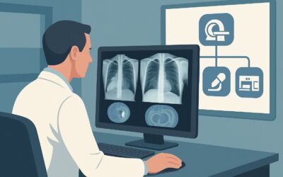
The ACR designates ‘Image-guided transthoracic needle biopsy’ as ‘Usually Appropriate’ because it directly addresses the need for a histologic diagnosis. This procedure, typically performed under CT guidance, allows for precise targeting of the mass while avoiding critical structures like the great vessels, heart, and airways. It provides a core needle sample suitable for histopathology, immunohistochemistry, and molecular studies, which are crucial for diagnosing and subtyping malignancies like lymphoma and lung cancer.
Rationale for Alternative Study Ratings:
- MRI chest without and with IV contrast (‘Usually Appropriate’): MRI is an excellent non-radiation alternative that offers superior soft-tissue contrast. It can be particularly valuable for assessing potential invasion of adjacent structures (e.g., chest wall, great vessels) or for characterizing posterior mediastinal masses suspected to be neurogenic. However, like PET/CT, it often cannot provide a definitive histologic diagnosis, and biopsy is frequently still required.
- CT chest with IV contrast (‘May be appropriate’): The patient has already had a CT as part of the PET/CT. A dedicated, contrast-enhanced CT might offer slightly better anatomic resolution or characterization of vascularity, but it is unlikely to resolve the diagnostic uncertainty. It adds another round of radiation (adult RRL ☢☢☢ 1-10 mSv) for what is often a low diagnostic yield at this stage.
- Radiography chest (‘Usually not appropriate’): A chest radiograph provides no additional information beyond the high-resolution CT and PET data already obtained and is therefore not helpful in this scenario.
What’s Next After Image-guided Transthoracic Needle Biopsy? Downstream Workflow
The results of the biopsy will dictate the subsequent clinical pathway. The goal of the procedure is to provide a definitive answer that breaks the diagnostic gridlock and allows for targeted treatment.
- If the Biopsy is Positive for Malignancy: This is the most direct outcome. A diagnosis of lymphoma, thymic carcinoma, metastatic lung cancer, or another malignancy will trigger a referral to the appropriate oncology service (medical, radiation, or surgical). The specific histology and molecular markers will guide the choice of chemotherapy, immunotherapy, radiation, or surgical resection.
- If the Biopsy Shows Granulomatous Inflammation: A finding of non-caseating granulomas would strongly suggest sarcoidosis, while caseating granulomas would point toward tuberculosis. This result shifts management away from oncology and toward pulmonology or infectious disease for further workup (e.g., cultures, ACE levels) and treatment with steroids or antimicrobial agents.
- If the Biopsy is Non-diagnostic: A non-diagnostic sample (e.g., insufficient tissue, crush artifact, necrosis) presents a challenge. The decision must be made whether to re-biopsy (perhaps with a larger core needle or a different approach) or to proceed to a more invasive surgical procedure like mediastinoscopy or video-assisted thoracoscopic surgery (VATS) for an excisional biopsy. The choice depends on the clinical suspicion for malignancy, the patient’s fitness for surgery, and the accessibility of the mass.
Pitfalls to Avoid (and When to Get Help)
Navigating the workup of an indeterminate mediastinal mass requires careful consideration to avoid common errors. One major pitfall is performing serial, inconclusive imaging studies when a tissue diagnosis is clearly needed. After an indeterminate PET/CT, another anatomic imaging study is unlikely to be definitive. Another common error is failing to provide adequate clinical history to the interventional radiologist and pathologist, as this context is crucial for planning the biopsy and interpreting the results. Finally, for masses adjacent to major vessels or the heart, ensure the biopsy is performed at a center with experienced interventional radiologists comfortable with complex thoracic procedures. If a percutaneous approach is deemed too high-risk, immediate escalation to a thoracic surgeon for a surgical biopsy should be considered.
Related ACR Topics and Tools
This article covers one specific scenario within the broader topic of mediastinal masses. For a comprehensive overview of all related clinical variants, from initial discovery to post-treatment follow-up, please consult our parent guide. Additional GigHz resources can help you apply these criteria in your daily practice.
- Parent Topic Hub: For breadth across all scenarios in Imaging of Mediastinal Masses, see our parent guide: Imaging of Mediastinal Masses: ACR Appropriateness Decoded.
- ACR Criteria Lookup: For adjacent scenarios not covered here, use the ACR Appropriateness Criteria Lookup.
- Imaging Protocols: For detailed techniques on recommended studies, explore the Imaging Protocol Library.
- Dose Conversations: To discuss cumulative exposure with patients, reference the Radiation Dose Calculator.
Frequently Asked Questions
Why not just proceed to surgical resection instead of a needle biopsy?
Surgical resection is a valid option, especially for a well-defined, solitary mass that appears resectable. However, for many conditions like lymphoma or sarcoidosis, the primary treatment is medical, not surgical. A pre-operative needle biopsy provides a definitive diagnosis that can prevent a major operation if the condition is best managed with chemotherapy or steroids.
Is MRI a good alternative to a biopsy in this scenario?
MRI is rated ‘Usually Appropriate’ and is an excellent problem-solving tool, especially for posterior mediastinal masses or to assess vascular invasion without radiation. However, its primary limitation is that it still provides a non-histologic, imaging-based diagnosis. If the PET/CT was already indeterminate, MRI may also be inconclusive, and a biopsy will likely still be required to guide treatment.
What are the main risks of a transthoracic needle biopsy for a mediastinal mass?
The most common risk is pneumothorax (collapsed lung), which occurs in a significant minority of cases but only requires a chest tube in a small fraction. Other risks include bleeding (hemoptysis or hemothorax) and, very rarely, air embolism. These risks are minimized when the procedure is performed by an experienced interventional radiologist using careful technique and planning.
If the mass is not FDG-avid on PET/CT, does that rule out malignancy?
No. While high FDG-avidity is sensitive for many aggressive malignancies, some tumors are not metabolically active or are too small to be resolved by PET. These include low-grade tumors like carcinoid tumors, some well-differentiated neuroendocrine tumors, and certain types of thymoma. A non-avid but otherwise suspicious mass on CT may still require a biopsy.
What if the mediastinal mass is not safely accessible for a percutaneous biopsy?
If the mass is surrounded by major blood vessels, the heart, or critical airways, a percutaneous transthoracic approach may be too risky. In these cases, alternative methods for obtaining tissue are considered. These include endobronchial ultrasound (EBUS)-guided biopsy if the mass is adjacent to the airway, or a surgical approach like mediastinoscopy or VATS for a direct excisional biopsy.
Reviewed by Pouyan Golshani, MD, Interventional Radiologist — May 29, 2026