Should You Order Imaging for an Uncomplicated COPD Exacerbation? An ACR Workflow
A 68-year-old male with a known history of Chronic Obstructive Pulmonary Disease (COPD) presents to your clinic with a three-day history of increased shortness of breath and a more productive cough. His vital signs are stable, and he has no fever or pleuritic chest pain. His oxygen saturation is 91% on room air, a slight dip from his baseline. You’ve initiated standard treatment with bronchodilators and systemic steroids, but the core clinical question remains: is imaging necessary to evaluate this seemingly uncomplicated exacerbation? This article provides a detailed workflow for this specific scenario, guided by the American College of Radiology (ACR) Appropriateness Criteria. For an immunocompetent adult with an uncomplicated acute COPD exacerbation, the ACR finds that a `Radiography chest` is Usually appropriate as the initial imaging study.
Who Fits the Uncomplicated Acute COPD Exacerbation Scenario?
This clinical workflow is designed for a specific patient population: an immunocompetent adult with a known diagnosis of COPD who presents with symptoms consistent with an acute exacerbation. These symptoms typically include an increase in dyspnea, sputum volume, or sputum purulence. The key qualifier is “uncomplicated.” This implies the patient is hemodynamically stable, afebrile or has a low-grade fever, and lacks new, alarming findings on physical examination, such as focal consolidation, signs of respiratory muscle fatigue, or altered mental status.
This guidance should not be applied to patients who fall outside these parameters. For instance, a patient presenting with high fever, hypotension, or severe respiratory distress may have a complicated infection or sepsis, requiring a different and more urgent diagnostic approach. Similarly, a patient with new, focal crackles on lung auscultation and pleuritic chest pain may better fit the ACR variant for pneumonia complicated by a suspected parapneumonic effusion. This focused workflow is for the common, low-acuity COPD flare where the primary goal of imaging is to rule out a simple, treatable comorbidity.
What Diagnoses Are You Working Up in an Uncomplicated COPD Flare?
When ordering a chest radiograph for an uncomplicated COPD exacerbation, the goal is to identify or exclude a limited set of common and consequential conditions that can mimic or complicate the presentation. The imaging study is not for confirming the exacerbation itself, which is a clinical diagnosis, but for assessing for alternative or co-existing pathologies.
The most common diagnosis being considered is a new community-acquired pneumonia. An underlying bacterial pneumonia can be a primary trigger for a COPD exacerbation, and its presence often alters management, specifically the decision to use antibiotics. A chest radiograph is highly effective at identifying a new infiltrate or consolidation that may not be apparent on physical examination, especially in patients with noisy baseline breath sounds from their underlying COPD.
A less common but critical consideration is a pneumothorax. Patients with COPD, particularly those with bullous emphysema, are at an increased risk for spontaneous pneumothorax. The acute onset of dyspnea can be indistinguishable from a typical exacerbation, making imaging essential for detection. A chest radiograph is the definitive initial study for this diagnosis.
Finally, imaging helps evaluate for signs of acute decompensated heart failure. The clinical syndromes of a COPD flare and a heart failure exacerbation can overlap significantly. A chest radiograph can reveal key findings such as cardiomegaly, pulmonary edema, Kerley B lines, or pleural effusions that would shift the diagnosis and management toward a cardiac etiology.
Why Is a Chest Radiograph the Recommended Study for This Presentation?
The American College of Radiology designates `Radiography chest` as Usually appropriate for an uncomplicated acute COPD exacerbation because it provides an excellent balance of diagnostic utility, accessibility, low cost, and minimal radiation exposure. It is sufficiently sensitive and specific for identifying the most common and critical differential diagnoses in this setting: pneumonia, significant pleural effusion, pneumothorax, and overt signs of congestive heart failure. For a patient who is clinically stable, the chest radiograph effectively answers the primary clinical question: is there a complicating factor that requires a change in management?
In contrast, more advanced imaging modalities are rated as Usually not appropriate for this specific initial workup. A `CT chest without IV contrast`, for example, offers much higher spatial resolution and is more sensitive for subtle infiltrates or small pneumothoraces. However, in an uncomplicated exacerbation, this increased sensitivity rarely changes initial management and comes at the cost of significantly higher radiation dose (☢☢☢ 1-10 mSv for CT versus ☢ <0.1 mSv for radiography) and expense. The ACR guidance suggests that the potential for over-diagnosis of clinically insignificant findings and the increased radiation burden outweigh the benefits in this low-acuity scenario.
Similarly, `CTA chest with IV contrast` is Usually not appropriate as a first-line study. While it is the gold standard for diagnosing pulmonary embolism (PE), PE is not typically suspected in an uncomplicated exacerbation without specific signs or symptoms like pleuritic chest pain, hemoptysis, or unexplained tachycardia. Ordering a CTA without clinical suspicion exposes the patient to both radiation and iodinated contrast unnecessarily. The principle is to match the intensity of the imaging workup to the clinical severity and pre-test probability of disease.
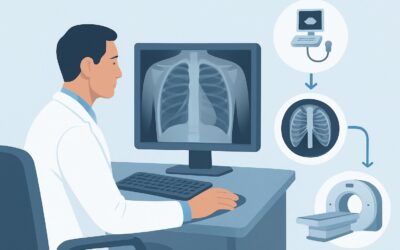
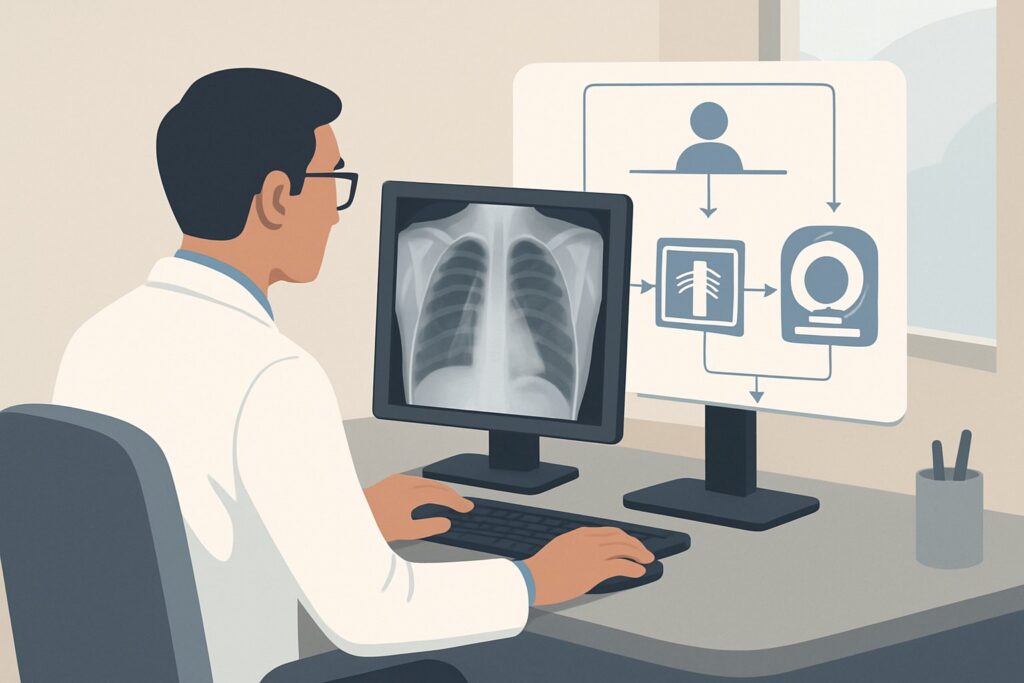
What’s Next After Radiography chest? Downstream Workflow
The results of the chest radiograph guide the subsequent clinical pathway. The workflow branches based on whether the findings are positive, negative, or indeterminate.
If the study is positive for a new infiltrate or consolidation, the diagnosis shifts to pneumonia complicating a COPD exacerbation. This finding typically solidifies the decision to initiate or continue antibiotic therapy targeted at common community-acquired pathogens. The patient’s disposition—whether they can be managed as an outpatient or require admission—will depend on their overall clinical stability, oxygen requirement, and other risk factors (e.g., CURB-65 score).
If the study is negative, showing no acute cardiopulmonary process, it reinforces the clinical diagnosis of an uncomplicated COPD exacerbation. Management continues to focus on bronchodilators, systemic corticosteroids, and supportive care. A negative radiograph provides reassurance that a significant pneumonia, pneumothorax, or heart failure is not the primary driver of the patient’s symptoms. If the patient fails to improve with standard therapy despite a negative chest x-ray, further investigation or a different diagnostic pathway may be considered.
If the study is indeterminate, such as showing hazy opacities that could represent atelectasis, scarring, or an early infiltrate, clinical correlation is paramount. The decision to treat with antibiotics may depend on other factors like sputum purulence and inflammatory markers. In rare cases where a significant finding is suspected but not confirmed, and the patient’s condition worsens, a follow-up chest radiograph or escalation to a non-contrast chest CT may be warranted.
Pitfalls to Avoid (and When to Get Help)
In managing this common scenario, several pitfalls can lead to diagnostic errors or unnecessary testing. A primary pitfall is over-reliance on a “normal” physical exam; patients with COPD often have diminished or noisy breath sounds that can obscure the classic signs of a new pneumonia. Another common error is ordering a CT scan as the initial imaging test for a stable, uncomplicated patient, leading to increased radiation exposure and cost without clear clinical benefit. Finally, be cautious not to attribute all worsening dyspnea to the COPD exacerbation itself; always consider mimics like pneumothorax and heart failure. If a patient’s symptoms are severe, atypical, or unresponsive to initial treatment, escalation to inpatient care or consultation with a pulmonologist is appropriate.
Related ACR Topics and Tools
For a comprehensive overview of imaging for respiratory illness and to explore related clinical scenarios, please refer to the resources below. These tools can help you select the right test for your specific patient and understand the technical details and radiation safety considerations for each study.
- For breadth across all scenarios in Acute Respiratory Illness in Immunocompetent Patients, see our parent guide: Acute Respiratory Illness in Immunocompetent Patients: ACR Appropriateness Decoded.
- To look up other clinical variants and see the full ACR guidelines, visit the Imaging Appropriateness Selector.
- For technical specifications on how imaging studies are performed, consult the Imaging Protocol Library.
- To discuss radiation dose with patients, use the Radiation Dose Calculator.
Frequently Asked Questions
Is a chest radiograph always necessary for a COPD exacerbation?
No, not always. For very mild exacerbations in reliable patients who are improving with initial therapy, clinical judgment may suggest deferring imaging. However, the ACR rates chest radiography as ‘Usually appropriate’ because it is a low-risk, high-value tool for ruling out common complicating factors like pneumonia, especially if the patient is being considered for hospital admission or is not responding as expected to treatment.
If the chest radiograph is negative but I still suspect a pulmonary embolism, what is the next step?
If there is a moderate to high clinical suspicion for pulmonary embolism (based on risk factors and clinical signs like tachycardia, pleuritic pain, or syncope), the appropriate next study is a CTA (Computed Tomography Angiography) of the chest. A negative chest radiograph does not rule out PE. The decision to order a CTA should be based on clinical scoring systems (like the Wells’ score) and/or D-dimer testing, not as a routine follow-up to a negative x-ray.
Why is chest CT ‘Usually not appropriate’ if it’s more sensitive than a radiograph?
While chest CT is more sensitive, its use in an uncomplicated, stable COPD exacerbation is generally not recommended because the increased diagnostic yield rarely changes initial management. It can lead to the detection of incidental, clinically insignificant findings, while exposing the patient to substantially more radiation and higher costs. The chest radiograph is sufficient to answer the key clinical questions in this low-acuity scenario.
Should I order a two-view (PA and lateral) or a single-view (portable AP) chest radiograph?
Whenever the patient’s condition permits, a two-view posteroanterior (PA) and lateral chest radiograph is preferred. This provides a more comprehensive evaluation of the lungs, heart, and mediastinum, and is better at localizing and characterizing abnormalities. A single-view anteroposterior (AP) portable radiograph is typically reserved for patients who are too ill or immobile to be transported to the radiology department.
Does this guidance apply to patients who are on chronic immunosuppressive therapy for other conditions?
No. This specific workflow is for immunocompetent patients. Patients on chronic steroids, chemotherapy, or other immunosuppressants are at risk for a wider range of opportunistic infections and may present with more subtle findings. Their workup is often more aggressive and may warrant earlier consideration of advanced imaging like CT, following a different ACR guideline.
Reviewed by Pouyan Golshani, MD, Interventional Radiologist — May 26, 2026