What Is the Best Initial Imaging for an Adult with Chronic Dyspnea of Unclear Etiology?
A 58-year-old patient presents to your clinic with a six-month history of progressive shortness of breath. The feeling is persistent, worsening with exertion, but the initial workup has been unrevealing. A cardiac evaluation, including an ECG and echocardiogram, was normal, and there are no clear signs of asthma or Chronic Obstructive Pulmonary Disease (COPD) on physical exam or spirometry. You suspect a non-cardiac, likely pulmonary, origin but the specific cause is unclear. The immediate clinical question is: what is the most appropriate first imaging study to order to begin clarifying the diagnosis?
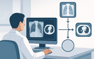
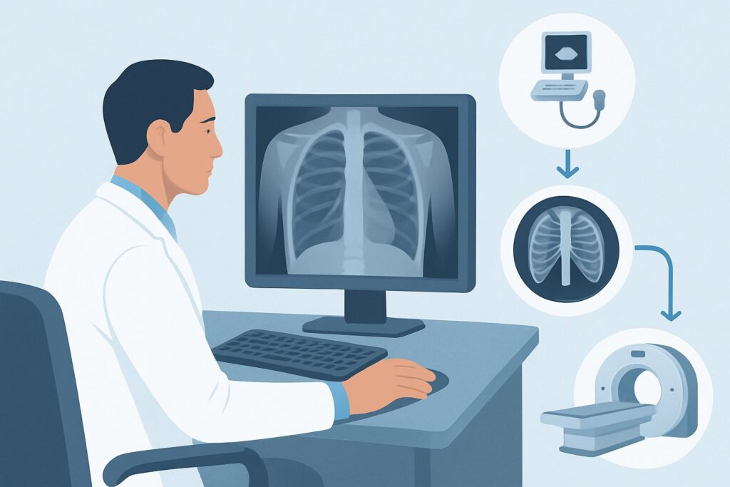
For this specific scenario—an adult with chronic dyspnea of unclear etiology requiring initial imaging—the American College of Radiology (ACR) Appropriateness Criteria rate a standard chest radiograph as Usually appropriate. This article provides a detailed workflow for this common clinical challenge, explaining the rationale for this choice, the differential diagnoses you are considering, and the critical downstream steps based on the initial imaging results.
Who Fits This Clinical Scenario?
This guidance applies specifically to adult patients presenting with chronic dyspnea, defined as shortness of breath lasting more than four weeks. The key feature of this scenario is the “unclear etiology.” This means that after a thorough history, physical examination, and initial non-imaging diagnostics (like basic labs, ECG, and possibly spirometry), a clear cause has not been identified. The patient does not have obvious signs or symptoms pointing to a specific diagnosis like heart failure, severe anemia, or a classic obstructive lung disease pattern.
This workflow is distinct from several related clinical situations. It does not apply if:
- COPD is strongly suspected: If the patient has a significant smoking history and clinical findings highly suggestive of COPD, the imaging workup follows a different path.
- Small airways disease is the primary concern: Patients with suspected constrictive bronchiolitis or similar conditions may require a different imaging approach, often involving expiratory CT imaging.
- There is a known history of prior COVID-19 infection: Post-COVID dyspnea has its own set of considerations, including the possibility of organizing pneumonia or fibrotic changes.
- Diaphragm dysfunction is suspected: If there are clinical clues pointing to diaphragmatic weakness or paralysis (e.g., orthopnea, paradoxical abdominal movement), the workup would include specialized tests like fluoroscopy (“sniff test”).
This article is for the common, undifferentiated patient where the goal of initial imaging is to narrow a broad differential diagnosis.
What Diagnoses Are You Working Up in This Scenario?
When ordering the initial imaging for undifferentiated chronic dyspnea, you are casting a wide but targeted net. The chest radiograph is intended to identify or exclude several key categories of pulmonary and thoracic disease.
Interstitial Lung Disease (ILD): This is a large group of disorders that cause progressive scarring of lung tissue. While a definitive diagnosis often requires high-resolution CT (HRCT), a standard chest radiograph can reveal tell-tale signs like reticular patterns, honeycombing, or volume loss, providing the first crucial clue. Idiopathic pulmonary fibrosis (IPF) is a key consideration in this category.
Occult Malignancy: A primary lung cancer or metastatic disease can present insidiously with dyspnea as the primary symptom. The chest radiograph is a highly effective screening tool for detecting pulmonary nodules, masses, lymphadenopathy, or malignant pleural effusions that would immediately redirect the diagnostic workup.
Chronic Infection or Inflammation: Conditions like smoldering tuberculosis, non-tuberculous mycobacterial infections, or chronic fungal pneumonia can manifest with dyspnea. Sarcoidosis, a systemic inflammatory disease, frequently involves the lungs and mediastinal lymph nodes, often producing characteristic findings of bilateral hilar adenopathy on a chest radiograph.
Pleural Disease: Chronic dyspnea can be caused by undiagnosed pleural effusions, pleural thickening, or plaques (e.g., from asbestos exposure). These abnormalities are typically well-visualized on a standard two-view chest radiograph, guiding further investigation or intervention.
Bronchiectasis: While severe cases are visible on radiographs as “tram tracking” or cystic changes, milder forms may be missed. However, a radiograph can suggest the diagnosis and prompt a confirmatory CT scan if clinical suspicion remains high.
Why Is a Chest Radiograph the Recommended Initial Study?
The ACR designates a standard posteroanterior (PA) and lateral chest radiograph as Usually appropriate for the initial imaging of an adult with chronic dyspnea of unclear etiology. This recommendation is based on a careful balance of diagnostic yield, safety, cost, and availability.
A chest radiograph serves as an excellent first-line examination. It is sensitive for detecting a wide range of significant parenchymal, pleural, and mediastinal abnormalities that cause chronic dyspnea. It can readily identify large masses, significant interstitial changes, cardiomegaly, large effusions, and hilar adenopathy. A normal chest radiograph is also a valuable finding, as it can lower the probability of many serious structural lung diseases and guide the clinician toward non-parenchymal causes like pulmonary vascular disease or neuromuscular weakness.
From a safety perspective, the radiation dose is minimal. A standard two-view chest radiograph delivers a very low effective dose (ACR Relative Radiation Level ☢, <0.1 mSv), which is a fraction of the dose from a CT scan and comparable to the background radiation a person receives over a few days.
In contrast, other more advanced imaging modalities are rated lower for this initial step:
- CT chest without IV contrast is rated May be appropriate (Disagreement). While more sensitive than a radiograph for subtle ILD or small nodules, the panel notes disagreement, reflecting that it is not always necessary as the first step. Jumping directly to CT exposes the patient to a significantly higher radiation dose (ACR RRL ☢☢☢, 1-10 mSv) and is less cost-effective when a simple radiograph can often guide the next steps appropriately.
- CT chest with IV contrast and MRI chest are rated Usually not appropriate. There is no initial indication for intravenous contrast when the primary question involves the lung parenchyma, and MRI has a very limited role in the general evaluation of chronic dyspnea due to its poor visualization of lung tissue.
- FDG-PET/CT is also Usually not appropriate as a first-line tool. It is a high-radiation (ACR RRL ☢☢☢☢, 10-30 mSv), high-cost functional imaging study reserved for specific indications like cancer staging or evaluating inflammatory activity, not for an initial, undifferentiated workup.
What’s Next After a Chest Radiograph? Downstream Workflow
The results of the initial chest radiograph are a critical branch point in the diagnostic algorithm. The subsequent steps depend entirely on whether the findings are positive, negative, or equivocal.
If the radiograph is positive: A finding of reticular opacities, honeycombing, or ground-glass changes strongly suggests ILD and should prompt a referral to a pulmonologist and a high-resolution CT (HRCT) of the chest for definitive characterization. If a mass, nodule, or significant lymphadenopathy is found, a contrast-enhanced CT of the chest is typically the next step to stage a potential malignancy and guide biopsy. The presence of a large pleural effusion would warrant a diagnostic and potentially therapeutic thoracentesis.
If the radiograph is negative: A normal chest radiograph is a common and important outcome. It significantly reduces the likelihood of advanced ILD, malignancy, or large-scale infection. However, it does not rule out all causes of dyspnea. The next steps should include a comprehensive set of pulmonary function tests (PFTs) to assess for obstructive or restrictive physiology not apparent on basic spirometry. If symptoms persist and PFTs are abnormal or inconclusive, this is the point where a non-contrast HRCT chest—the study rated May be appropriate—becomes a logical next step to look for subtle ILD or small airways disease missed on the radiograph.
If the radiograph is indeterminate: Vague or equivocal findings, such as subtle basal opacities or questionable hilar fullness, also warrant progression to a CT scan of the chest for clarification. A non-contrast CT is usually sufficient to clarify parenchymal questions, while a contrast-enhanced study may be needed if there is concern for a vascular or mediastinal abnormality.
Pitfalls to Avoid (and When to Get Help)
In this common clinical scenario, a few pitfalls can delay diagnosis or lead to unnecessary testing. First, avoid under-interpreting a “normal” chest radiograph in a patient with persistent or worsening symptoms, especially if they have risk factors like a history of smoking or autoimmune disease. Early ILD can be radiographically occult. Second, resist the urge to order a CT scan as the initial test for every patient; the chest radiograph is a powerful and sufficient triage tool in most cases. Third, ensure a high-quality, two-view (PA and lateral) study is obtained, as a single anteroposterior (AP) view can miss significant pathology.
If the chest radiograph is negative but the patient’s dyspnea continues unabated, or if PFTs reveal a significant abnormality (e.g., a reduced diffusion capacity for carbon monoxide, DLCO), it is time to escalate. This typically involves a consultation with a pulmonologist for further evaluation, which may include advanced imaging, bronchoscopy, or a cardiopulmonary exercise test.
Related ACR Topics and Tools
This article covers one specific scenario in depth. For a broader view of imaging for other causes of chronic shortness of breath, or to explore the tools used to make these decisions, the following resources are valuable.
- For breadth across all scenarios in Chronic Dyspnea-Noncardiovascular Origin, see our parent guide: Chronic Dyspnea-Noncardiovascular Origin: ACR Appropriateness Decoded.
- To look up other clinical scenarios, use the ACR Appropriateness Criteria Lookup.
- For details on imaging techniques, consult the Imaging Protocol Library.
- To discuss radiation exposure with patients, the Radiation Dose Calculator can help frame the risks.
Frequently Asked Questions
Why not start with a high-resolution CT (HRCT) if it’s more sensitive than a chest radiograph?
While an HRCT is more sensitive for subtle interstitial lung disease, it comes with a significantly higher radiation dose and cost. The ACR recommends a stepwise approach where the chest radiograph acts as a highly effective triage tool. It can confirm a diagnosis or guide the need for CT in many cases, avoiding unnecessary radiation and expense for patients whose radiograph is normal and whose symptoms may resolve or be explained by non-parenchymal causes.
If the chest radiograph is normal, can I confidently rule out serious lung disease?
No. A normal chest radiograph significantly lowers the probability of many advanced lung diseases like extensive fibrosis, large tumors, or sarcoidosis, but it cannot definitively rule out all pathology. Early interstitial lung disease, small airways disease (like constrictive bronchiolitis), and small nodules can be missed. If clinical suspicion remains high despite a normal radiograph, further testing like full pulmonary function tests (PFTs) and potentially a CT scan is warranted.
Is a single AP portable chest x-ray sufficient for this initial workup?
A single anteroposterior (AP) portable view is not ideal and should be avoided if the patient is ambulatory. The standard of care is a two-view study consisting of a posteroanterior (PA) and a lateral view. The lateral view is critical for evaluating the retrosternal and retrocardiac spaces, the posterior costophrenic sulci, and the thoracic spine, areas where pathology can be hidden on a single frontal view.
When should I add IV contrast to a follow-up CT scan?
Intravenous contrast is generally not needed if the primary question is about the lung parenchyma (e.g., evaluating for interstitial lung disease or bronchiectasis). A contrast-enhanced CT is indicated if the chest radiograph or clinical picture suggests a mediastinal mass, hilar adenopathy, a suspected vascular issue like a pulmonary embolism (though CT angiography protocol would be used), or a suspected malignancy that requires staging.
Does this guidance apply to patients with acute dyspnea?
No, this guidance is specifically for chronic dyspnea, which is defined as lasting four weeks or more. The differential diagnosis and imaging workup for acute dyspnea are different and are focused on emergent conditions like pulmonary embolism, pneumonia, pneumothorax, and acute heart failure exacerbation. That scenario is covered under different ACR Appropriateness Criteria topics.
Reviewed by Pouyan Golshani, MD, Interventional Radiologist — May 29, 2026