

Your Patient Has an SMA Embolus and Peritonitis: What’s the ACR-Guided Next Step?
ACR-guided imaging workflow for Recent onset abdominal pain, peritoneal signs, and known atrial fibrillation. CTA shows filling defect in the proximal SMA
ACR-guided imaging workflow for Recent onset abdominal pain, peritoneal signs, and known atrial fibrillation. CTA shows filling defect in the proximal SMA

ACR-guided imaging workflow for Recent onset abdominal pain, no peritoneal signs, and known atrial fibrillation. CTA shows calcified atherosclerotic plaque

ACR-guided imaging workflow for Recent onset abdominal pain, no peritoneal signs, and known atrial fibrillation. CTA shows filling defect in proximal SMA c

ACR-guided imaging workflow for Suspected soft tissue infection. Initial radiographs show soft tissue gas (without puncture wound) or are normal with high

ACR-guided imaging workflow for Suspected soft tissue infection. History of puncture wound with possible retained foreign body. Radiographs normal. Next im

ACR-guided imaging workflow for Suspected septic arthritis with arthroplasty or other implanted intra-articular surgical hardware. Initial radiographs norm

ACR-guided imaging workflow for Suspected osteomyelitis or soft tissue infection with implanted extra-articular surgical hardware. Initial radiographs norm
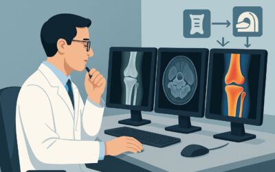
ACR-guided imaging workflow for Suspected osteomyelitis. Initial radiographs normal or with findings suggestive of osteomyelitis. Next imaging study. (Susp

ACR-guided imaging workflow for Suspected septic arthritis or soft tissue infection. Initial radiographs normal or with findings suggestive of joint effusi

ACR-guided imaging workflow for Chronic elbow pain. Suspect chronic epicondylalgia or tendon tear. Refractory to empirical treatment. Radiographs normal or