What Imaging Is Best for Suspected Osteomyelitis After Normal or Equivocal Radiographs?
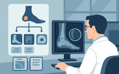
A 10-year-old boy presents to the emergency department with a two-week history of worsening, localized pain in his distal tibia and a low-grade fever. He denies any specific trauma. You obtain initial radiographs of the leg, which show only subtle soft tissue swelling and no definitive fracture or periosteal reaction. Your clinical suspicion for osteomyelitis remains high, but the x-ray is inconclusive. What is the next, most appropriate imaging study to confirm or exclude the diagnosis and guide management? This is a common clinical crossroads where choosing the right advanced imaging is critical for timely treatment.
According to the American College of Radiology (ACR) Appropriateness Criteria, for a patient with suspected osteomyelitis where initial radiographs are normal or have suggestive findings, an MRI of the area of interest without and with IV contrast is rated as Usually Appropriate.
Who Fits This Clinical Scenario?
This guidance is specifically for patients where there is a clinical concern for osteomyelitis of the appendicular skeleton or pelvis, and the initial step of obtaining radiographs has already been completed.
Inclusion criteria for this workflow:
- Focal bone pain, tenderness, or swelling.
- Systemic signs of infection may be present (fever, elevated inflammatory markers like C-reactive protein or erythrocyte sedimentation rate).
- Initial radiographs are either completely normal or show non-specific, equivocal findings such as deep soft tissue swelling, periosteal reaction, or subtle lucency.
Exclusion criteria (these patients follow different guidelines):
- Suspected Septic Arthritis: If the primary concern is infection within a joint, characterized by painful effusion and limited range of motion, the imaging workup prioritizes evaluating the joint space and synovium.
- Presence of Surgical Hardware: Patients with plates, screws, or other extra-articular hardware require specific imaging protocols (often involving metal artifact reduction sequences on MRI or specialized nuclear medicine scans) to evaluate for hardware-associated infection. This is a distinct ACR scenario.
- Diabetic Foot Infection: This is a complex condition involving neuropathy, vascular compromise, and often polymicrobial infection. The ACR has separate, dedicated guidelines for imaging the diabetic foot, which differs significantly from this workflow.
- Suspected Spinal Osteomyelitis (Discitis-osteomyelitis): Infection of the spine has its own unique diagnostic pathway, and this guidance does not apply.
What Diagnoses Are You Working Up in This Scenario?
When ordering advanced imaging after an inconclusive radiograph, you are evaluating a differential that extends beyond simple infection. The goal is to differentiate osteomyelitis from its mimics, which can present with similar clinical and radiographic signs.
Acute Osteomyelitis
This is the primary diagnosis of concern. Bacteria, most commonly Staphylococcus aureus, seed the bone marrow, leading to an inflammatory response, edema, and eventually bone destruction. MRI is exceptionally sensitive to the early marrow edema that occurs days before any changes are visible on radiographs. Contrast enhancement helps identify abscess formation, which is a critical finding for determining the need for surgical drainage.
Stress Fracture or Other Occult Trauma
In active children, adolescents, or adults who are athletes or laborers, a stress fracture can present with focal bone pain and tenderness. Initial radiographs are often normal. MRI can show marrow edema and a fracture line, effectively distinguishing it from the more diffuse, ill-defined edema and potential abscess seen in osteomyelitis.
Primary Bone Tumor
Less common but critical to diagnose, certain malignancies can mimic osteomyelitis. In children and young adults, Ewing sarcoma can present with pain, fever, and periosteal reaction. In older adults, lymphoma or metastatic disease can manifest similarly. MRI is crucial for characterizing the lesion, assessing for a soft tissue mass, and determining the extent of disease, which are key features that help differentiate tumor from infection.
Cellulitis or Pyomyositis
A severe soft tissue infection adjacent to the bone can cause reactive marrow edema and periosteal inflammation, mimicking osteomyelitis on clinical exam and even on imaging. MRI excels at delineating the primary site of infection. Identifying an intramuscular abscess (pyomyositis) or diffuse cellulitis without direct marrow involvement changes management from prolonged bone-penetrating antibiotics to a focus on soft tissue treatment.
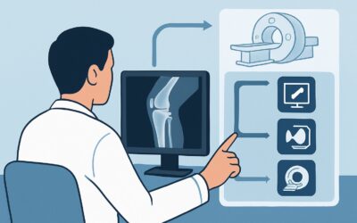
Why Is MRI Without and With IV Contrast the Recommended Study?
The ACR rates MRI of the area of interest without and with IV contrast as Usually Appropriate because of its superior soft tissue contrast, high sensitivity for early disease, and lack of ionizing radiation. It directly visualizes the pathophysiology of osteomyelitis.
The rationale for this recommendation includes:
- High Sensitivity and Specificity: MRI can detect the increased water content of marrow edema within hours to days of infection onset, long before the 10-14 days it typically takes for bone destruction to become apparent on radiographs. This makes it the most sensitive modality for early diagnosis. When combined with clinical findings, its specificity is also very high.
- Anatomic Detail: MRI provides a detailed map of the infection. It can precisely locate an intramedullary abscess, identify a sinus tract extending to the skin, show an adjacent septic joint effusion, and define the extent of soft tissue involvement. This information is invaluable for surgical planning.
- Role of IV Contrast: While a non-contrast MRI (also rated Usually Appropriate) is excellent for detecting marrow edema, the addition of a gadolinium-based contrast agent significantly improves diagnostic confidence. Contrast helps to:
- Delineate abscess cavities, which typically show a peripherally enhancing rim.
- Distinguish between viable, enhancing tissue and non-viable, necrotic tissue (sequestrum).
- Assess the degree of soft tissue inflammation and vascularity.
- Safety Profile: MRI avoids the use of ionizing radiation (0 mSv), which is a particularly important consideration in pediatric and young adult patients who may require follow-up imaging.
Why are other studies rated lower for this specific scenario?
- Three-Phase Bone Scan: Rated May be appropriate. While highly sensitive for detecting areas of increased bone turnover, it is not specific. A “hot spot” can be caused by infection, fracture, tumor, or arthritis. Its poor spatial resolution makes it difficult to distinguish bone from soft tissue infection or to identify an abscess.
- CT with IV Contrast: Rated May be appropriate. CT is excellent for evaluating cortical bone destruction, periosteal reaction, and identifying a sequestrum (a piece of dead bone). However, it is insensitive to the early marrow changes of osteomyelitis, making it a poor choice for early diagnosis when radiographs are normal. It also involves a variable but significant radiation dose.
What’s Next After MRI? Downstream Workflow
The results of the MRI will guide your next steps, creating a clear decision tree for patient management.
- If the MRI is positive for osteomyelitis: This confirms the diagnosis. The next step is typically consultation with orthopedic surgery and infectious disease specialists. A bone biopsy, often performed under image guidance, may be necessary to obtain cultures to tailor antibiotic therapy. The MRI findings will also determine if surgical debridement is required, especially in the presence of a significant abscess or sequestrum.
- If the MRI is negative for osteomyelitis: A technically adequate negative MRI has a very high negative predictive value and effectively rules out osteomyelitis. The clinical workup should pivot to the other differential diagnoses. If a stress fracture is identified, management involves rest and activity modification. If a tumor is suspected, an orthopedic oncology consultation and biopsy are warranted. If only soft tissue inflammation is seen, treatment can be focused on cellulitis or myositis.
- If the MRI is indeterminate: In some cases, findings like non-specific marrow edema may be equivocal. The next step depends on the clinical context. If suspicion for infection remains high, a short-interval follow-up MRI in 1-2 weeks may show evolution. Alternatively, in complex cases, a more functionally-oriented study like an FDG-PET/CT (rated May be appropriate) could be considered to assess for metabolic activity suggestive of infection or malignancy. A biopsy remains the ultimate problem-solver for indeterminate findings.
Pitfalls to Avoid (and When to Get Help)
Navigating the workup for suspected osteomyelitis requires avoiding several common pitfalls that can delay diagnosis or lead to misinterpretation.
1. False Reassurance from a Normal Radiograph: The most common error is stopping the workup after a normal x-ray in a patient with high clinical suspicion. Remember that radiographic changes lag behind the infectious process by up to two weeks.
2. Omitting IV Contrast on the MRI: While a non-contrast study is still valuable, forgoing contrast can mean missing a drainable abscess. Unless the patient has a severe contraindication, ordering the MRI “without and with contrast” provides the most complete diagnostic picture.
3. Over-reliance on Inflammatory Markers: While helpful, ESR and CRP are non-specific. They can be elevated in trauma, malignancy, and autoimmune conditions. They support a diagnosis of osteomyelitis but do not make it.
4. Misinterpreting Reactive Edema: Remember that marrow edema can be a reactive change from an overlying soft tissue infection or adjacent septic arthritis. Carefully scrutinize the entire imaging volume to find the primary source of pathology.
If the MRI findings are complex, equivocal, or discordant with the clinical picture, a consultation with a musculoskeletal radiologist is essential to help interpret the findings and suggest the most appropriate next steps.
Related ACR Topics and Tools
This article focuses on a single clinical scenario. For a comprehensive overview of all related presentations and for tools to help you in your practice, please refer to the following resources.
For breadth across all scenarios in Suspected Osteomyelitis, Septic Arthritis, or Soft Tissue Infection (Excluding Spine and Diabetic Foot), see our parent guide: Suspected Osteomyelitis, Septic Arthritis, or Soft Tissue Infection (Excluding Spine and Diabetic Foot): ACR Appropriateness Decoded.
- ACR Appropriateness Criteria Lookup — for adjacent scenarios
- Imaging Protocol Library — for technique on the recommended study
- Radiation Dose Calculator — for cumulative dose conversations
Frequently Asked Questions
Why not just get a CT scan if the x-ray is normal for suspected osteomyelitis?
CT is rated as ‘May be appropriate’ but is not the preferred next step. While excellent for showing cortical bone destruction and sequestra, CT is insensitive to the early changes of osteomyelitis within the bone marrow. MRI can detect marrow edema days to weeks before any changes are visible on CT, making it far superior for early diagnosis.
Is an MRI without contrast good enough for suspected osteomyelitis?
A non-contrast MRI is also rated ‘Usually Appropriate’ by the ACR and is highly sensitive for detecting the marrow edema characteristic of early osteomyelitis. However, the addition of IV gadolinium contrast is strongly recommended because it significantly improves the ability to identify and delineate abscesses, which is critical information for determining the need for surgical drainage.
How long does it take for radiographs to become positive for osteomyelitis?
Radiographic evidence of osteomyelitis, such as periosteal reaction, cortical erosion, or lucency, typically lags behind the onset of infection by 10 to 14 days. This delay is why advanced imaging like MRI is crucial when clinical suspicion is high despite normal or equivocal initial x-rays.
What if my patient has a pacemaker or other contraindication to MRI?
If a patient has an absolute contraindication to MRI, you must turn to the ‘May be appropriate’ alternatives. A three-phase nuclear medicine bone scan is a sensitive option, though not specific. If available, a labeled white blood cell (WBC) scan can offer more specificity for infection. CT with IV contrast can also be used to look for later-stage findings like bone destruction or abscess.
Does this guidance apply to patients with diabetic foot ulcers?
No. Suspected osteomyelitis in the setting of a diabetic foot ulcer is a distinct and complex clinical scenario. The ACR has separate, specific Appropriateness Criteria for this condition, as the diagnostic challenges involve underlying neuropathy, peripheral vascular disease, and potential for soft tissue ulcers to obscure the exam. This article’s guidance should not be applied to that patient population.
Reviewed by Pouyan Golshani, MD, Interventional Radiologist — May 30, 2026