What Imaging Is Next for Suspected Septic Arthritis with Normal Radiographs?
A 45-year-old man presents to the emergency department with a three-day history of a severely painful, swollen, and erythematous right knee. He has a low-grade fever and his C-reactive protein is markedly elevated. You obtain initial radiographs of the knee, which demonstrate a moderate joint effusion and soft tissue swelling, but no fracture, dislocation, or destructive osseous changes. The clinical suspicion for septic arthritis is high, but you need to confirm the diagnosis, rule out mimics like a deep abscess, and potentially guide aspiration. What is the most appropriate next imaging study to order in this urgent clinical setting?
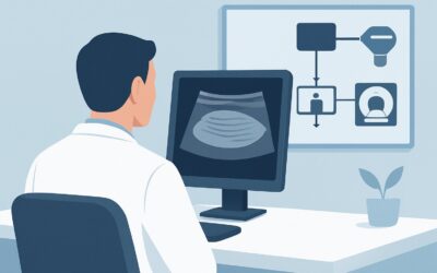
This article provides a detailed workflow for this specific scenario, grounded in the American College of Radiology (ACR) Appropriateness Criteria. For a patient with suspected septic arthritis or soft tissue infection whose initial radiographs are non-diagnostic, ‘US area of interest’ is rated Usually Appropriate as the next imaging step.
Who Fits This Clinical Scenario for Suspected Joint Infection?
This guidance applies to a well-defined patient population, typically adults or children presenting with acute signs of a focal musculoskeletal infection. The key inclusion criteria are:
- Clinical suspicion for septic arthritis (e.g., acute monoarticular pain, erythema, warmth, limited range of motion) or a significant soft tissue infection (e.g., suspected abscess).
- Systemic signs of infection, such as fever or elevated inflammatory markers (leukocytosis, high ESR/CRP), are often present but not required.
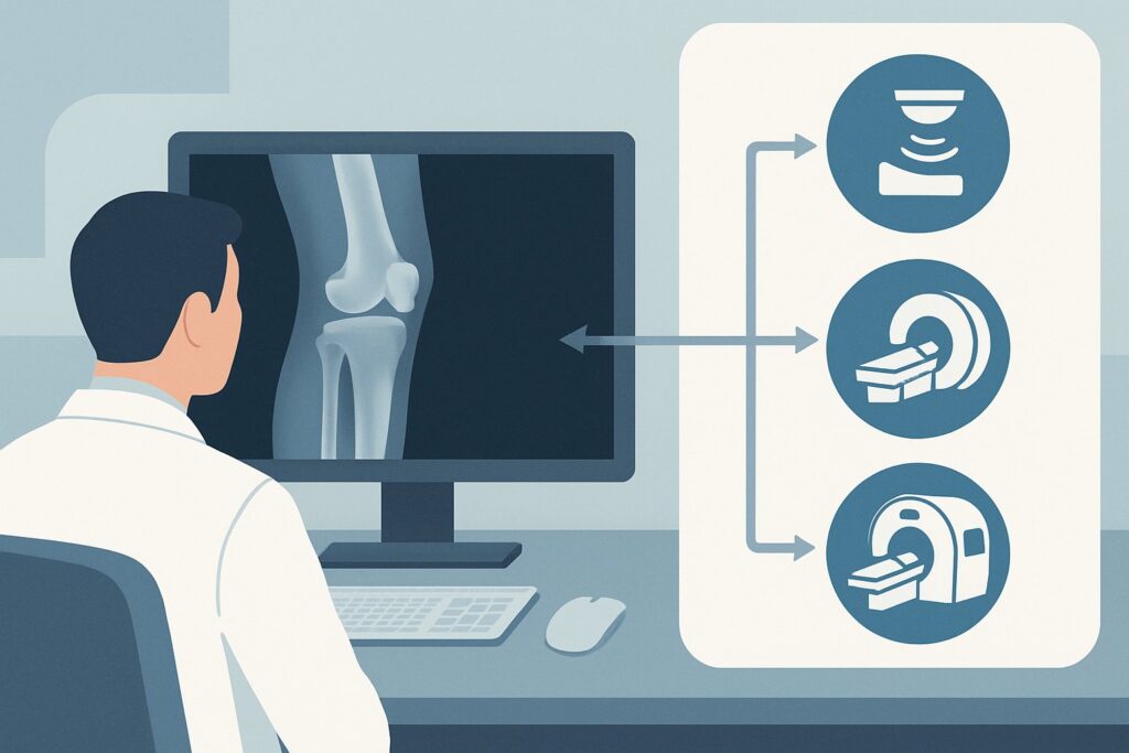
- Initial radiographs of the area have already been performed and are either entirely normal or show only non-specific findings like soft tissue swelling or a joint effusion.
It is critical to distinguish this presentation from similar but distinct clinical situations that follow different diagnostic pathways. This workflow does not apply if:
- Osteomyelitis is strongly suspected or seen on radiographs. If initial X-rays show cortical destruction, periosteal reaction, or other signs of bone infection, the workup follows the suspected osteomyelitis variant.
- Surgical hardware is present. Patients with joint replacements or other intra-articular hardware require a specialized workup for prosthetic joint infection.
- A retained foreign body is the primary concern. Following a puncture wound, the imaging focus shifts to identifying materials like wood or plastic, which may require different modalities.
- The patient has a diabetic foot ulcer. This is a complex condition with its own dedicated ACR Appropriateness Criteria.
What Diagnoses Are You Working Up in This Scenario?
When a patient presents with an acutely inflamed joint and non-diagnostic radiographs, the differential diagnosis is focused on conditions requiring urgent intervention. The choice of imaging is driven by the need to distinguish between these possibilities.
Septic Arthritis
This is the most urgent diagnosis to confirm or exclude. A bacterial infection within the synovial space is an orthopedic emergency, as enzymatic degradation can destroy articular cartilage within 24-48 hours, leading to permanent joint damage. Imaging is used to identify a joint effusion, assess synovial thickening and hyperemia, and, most importantly, guide arthrocentesis for definitive diagnosis via fluid analysis and culture.
Soft Tissue Abscess or Cellulitis
A deep soft tissue infection adjacent to a joint can clinically mimic septic arthritis, causing what is known as a “sympathetic effusion.” Differentiating a drainable fluid collection (abscess) from diffuse inflammation (cellulitis) is critical for management. While cellulitis is treated with antibiotics alone, an abscess requires drainage. Ultrasound excels at this distinction.
Inflammatory Arthropathy (e.g., Gout, Pseudogout)
An acute flare of a crystal-induced arthropathy like gout or pseudogout can be clinically indistinguishable from septic arthritis, presenting with severe pain, swelling, and erythema. While joint aspiration is the gold standard for diagnosis (identifying crystals), imaging can help locate the optimal site for aspiration, especially in complex joints.
Bursitis
Inflammation or infection of a bursa (e.g., prepatellar or olecranon bursitis) can also present with focal swelling and pain. Imaging can confirm that the fluid and inflammation are confined to the bursa and have not extended into the adjacent joint space, which would significantly alter management.
Why Is Ultrasound of the Area of Interest the Recommended Next Study?
For a patient with suspected septic arthritis or soft tissue infection and non-diagnostic initial radiographs, the ACR rates ‘US area of interest’ as Usually Appropriate. This recommendation is based on its high diagnostic utility, safety profile, and accessibility for this specific clinical question.
Ultrasound is highly sensitive for detecting joint effusions, even small ones that may be subtle on radiographs. It provides dynamic, real-time evaluation of the joint and surrounding soft tissues. A sonographer can identify synovial thickening and increased blood flow on color Doppler imaging, which are suggestive of inflammation or infection. Crucially, ultrasound can distinguish a simple effusion from a complex, debris-filled one (pyarthrosis) and can clearly differentiate an intra-articular process from an overlying cellulitis, abscess, or bursitis.
Perhaps its most significant advantage is its utility in guiding procedures. Ultrasound allows for real-time, image-guided aspiration of a joint effusion or abscess. This increases the success rate of the procedure, reduces patient discomfort, and minimizes the risk of a “dry tap,” ensuring a diagnostic fluid sample is obtained. Because it uses no ionizing radiation (0 mSv), it is safe for all patients, including children and pregnant women.
Why are other studies rated differently for this initial step?
- MRI without and with IV contrast is also rated Usually Appropriate and provides excellent anatomic detail of synovium, cartilage, bone marrow, and soft tissues. However, it is more expensive, less readily available in an emergency setting, and takes longer to perform than ultrasound. It is often reserved for cases where ultrasound is equivocal or when osteomyelitis is a strong concurrent concern.
- CT with IV contrast is also Usually Appropriate but is used less frequently as the primary next step. While it can identify fluid collections and rim-enhancing abscesses, it involves ionizing radiation (Varies) and offers inferior soft tissue contrast compared to MRI and less functional information than ultrasound. Its main role is in patients with contraindications to MRI.
- A 3-phase bone scan is rated Usually Not Appropriate for this scenario. It is sensitive for inflammation but highly non-specific; it cannot reliably distinguish between septic arthritis, cellulitis, and osteomyelitis. It also involves a significant radiation dose (☢☢☢ 1-10 mSv).
What’s Next After Ultrasound? Downstream Workflow
The results of the ultrasound directly guide the subsequent clinical and diagnostic steps. The workflow branches based on the key findings.
If the ultrasound is POSITIVE for a significant joint effusion:
The immediate next step is diagnostic and therapeutic arthrocentesis, which can be performed under ultrasound guidance. The synovial fluid should be sent for cell count with differential, Gram stain, culture, and crystal analysis. A positive Gram stain or a high synovial white blood cell count (typically >50,000/µL with neutrophil predominance) is highly suggestive of septic arthritis, prompting urgent surgical consultation for irrigation and debridement, along with empiric intravenous antibiotics.
If the ultrasound is POSITIVE for a drainable soft tissue fluid collection (abscess):
The next step is drainage, which can often be performed percutaneously with ultrasound guidance, along with antibiotic therapy. If the abscess is large, multiloculated, or in a surgically challenging location, a surgical consultation for incision and drainage is warranted.
If the ultrasound is NEGATIVE or shows only simple cellulitis/synovitis:
If no significant effusion or drainable collection is found, but clinical suspicion remains high, management depends on the clinical picture. The patient may be treated for cellulitis with antibiotics. If symptoms fail to improve or if there is a strong concern for underlying osteomyelitis that ultrasound cannot assess, the next appropriate step is often an MRI without and with IV contrast to evaluate the bone marrow and deep soft tissues in greater detail.
If the ultrasound is INDETERMINATE:
In cases where findings are equivocal (e.g., a complex but non-aspirated fluid collection, technically limited study), proceeding to MRI is the logical next step to provide a more definitive anatomic evaluation.
Pitfalls to Avoid (and When to Get Help)
Navigating the workup for suspected septic arthritis requires vigilance to avoid common diagnostic errors.
- Pitfall 1: Delaying Arthrocentesis. Time is cartilage. Once a joint effusion is identified on ultrasound in a patient with high clinical suspicion, do not delay aspiration pending other tests. Septic arthritis is a clinical diagnosis confirmed by synovial fluid analysis; imaging’s primary role is to confirm an effusion and guide the needle.
- Pitfall 2: Ascribing a “Negative” Ultrasound to a Lack of Infection. Ultrasound is operator-dependent. In a patient with ongoing high clinical suspicion but a non-diagnostic ultrasound, do not prematurely close the workup. Consider repeating the exam or escalating to MRI.
- Pitfall 3: Forgetting Crystal Arthropathy. Always send synovial fluid for crystal analysis. Septic arthritis and an acute gout flare can coexist, and failing to diagnose and treat gout can lead to recurrent, debilitating symptoms.
If a patient’s condition deteriorates despite initial treatment, or if imaging reveals extensive soft tissue necrosis or signs of necrotizing fasciitis (a rare but catastrophic mimic), immediate surgical consultation is mandatory.
Related ACR Topics and Tools
This article focuses on one specific clinical scenario. For a comprehensive overview of imaging for musculoskeletal infections and to explore related clinical variants, please consult the resources below.
- For breadth across all scenarios in Suspected Osteomyelitis, Septic Arthritis, or Soft Tissue Infection (Excluding Spine and Diabetic Foot), see our parent guide: Suspected Osteomyelitis, Septic Arthritis, or Soft Tissue Infection (Excluding Spine and Diabetic Foot): ACR Appropriateness Decoded.
- ACR Appropriateness Criteria Lookup — for adjacent scenarios
- Imaging Protocol Library — for technique on the recommended study
- Radiation Dose Calculator — for cumulative dose conversations
Frequently Asked Questions
Why not go straight to MRI for suspected septic arthritis?
While MRI is also rated ‘Usually Appropriate’ and provides excellent detail, ultrasound is often preferred as the *next* step after radiographs because it is faster, more accessible, less expensive, and involves no radiation. Most importantly, it allows for real-time guided aspiration of joint fluid, which is the definitive diagnostic step. MRI is typically reserved for cases where ultrasound is inconclusive or if there’s a strong suspicion of concurrent osteomyelitis.
Can ultrasound reliably distinguish septic arthritis from gout?
No, not definitively on its own. Both conditions can cause a joint effusion and synovial inflammation (hyperemia) visible on ultrasound. While some sonographic features like the ‘double contour sign’ are suggestive of gout, there is significant overlap. The primary role of ultrasound is to confirm an effusion and guide aspiration. The synovial fluid analysis is what differentiates infection from crystal arthropathy.
What if the patient has a contraindication to MRI, but the ultrasound is inconclusive?
If MRI is contraindicated (e.g., due to an incompatible implanted device or severe claustrophobia) and the ultrasound is non-diagnostic, ‘CT area of interest with IV contrast’ is rated ‘Usually Appropriate’ and becomes the next best option. CT can identify joint effusions, rim-enhancing fluid collections suggestive of an abscess, and cortical bone erosion, although it has lower soft tissue resolution than MRI.
Is an ultrasound necessary if a joint effusion is obvious on physical exam?
Even if an effusion is clinically obvious, ultrasound is still highly valuable. It confirms the effusion is intra-articular, characterizes the fluid (simple vs. complex), evaluates the synovium, and assesses the surrounding soft tissues for an abscess. Most importantly, it facilitates a safer and more successful needle aspiration, especially in deeper joints like the hip or in patients with obesity.
Does a normal ultrasound rule out septic arthritis?
A normal ultrasound that shows no joint effusion and no synovial inflammation makes septic arthritis highly unlikely. However, in the very early stages of infection, an effusion may not have accumulated yet. If clinical suspicion remains very high despite a normal initial ultrasound, the patient should be observed closely, and repeating the imaging in 12-24 hours or proceeding to MRI may be considered.
Reviewed by Pouyan Golshani, MD, Interventional Radiologist — May 30, 2026