

What’s the Best Initial Imaging for a Vaginal Bulge or Suspected Pelvic Organ Prolapse?
ACR-guided imaging workflow for Vaginal protrusion or bulge, or clinically suspected pelvic organ prolapse. Initial imaging. (Pelvic Floor Dysfunction in F
ACR-guided imaging workflow for Vaginal protrusion or bulge, or clinically suspected pelvic organ prolapse. Initial imaging. (Pelvic Floor Dysfunction in F
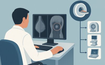
ACR-guided imaging workflow for Female. Suspicious axillary node on any other imaging modality (excluding mammography and ultrasound). Next imaging study.
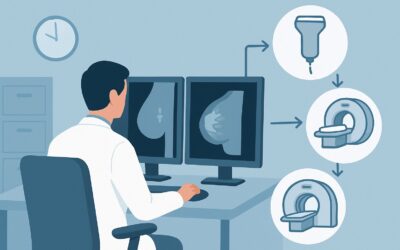
ACR-guided imaging workflow for Female. Suspicious axillary node on mammography or ultrasound. Next imaging study. (Imaging of the Axilla): recommended stu
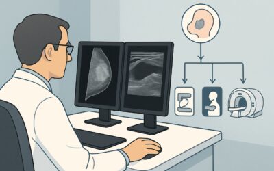
ACR-guided imaging workflow for Female. Newly diagnosed locally recurrent breast cancer. Initial imaging of the axilla following diagnostic mammography or

ACR-guided imaging workflow for Female. Breast cancer, greater than 2 cm in size, clinical node-positive. Imaging of the axilla after completion of neoadju
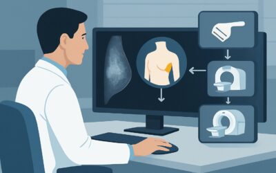
ACR-guided imaging workflow for Female. Breast cancer, greater than 2 cm in size, clinical node-negative. Imaging of the axilla after completion of neoadju
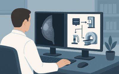
ACR-guided imaging workflow for Female. Breast cancer, greater than 2 cm in size, at mid-treatment of neoadjuvant chemotherapy, with initial clinical node-
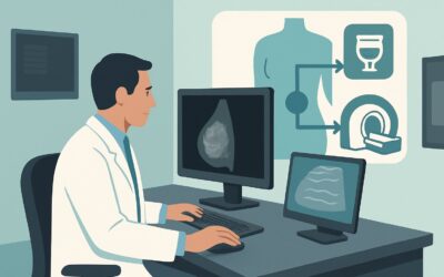
ACR-guided imaging workflow for Female. Newly diagnosed breast cancer, greater than 2 cm, with clinical node-positive. Initial imaging of the axilla follow
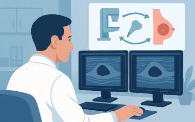
ACR-guided imaging workflow for Female. Newly diagnosed breast cancer, greater than 2 cm, with clinical node-negative. Initial imaging of the axilla follow
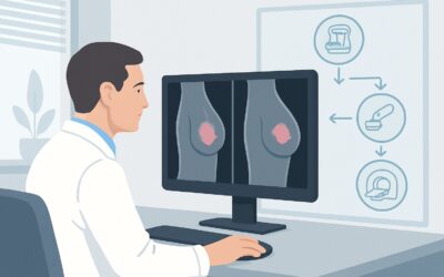
ACR-guided imaging workflow for Female. Newly diagnosed breast cancer, 2 cm or less, with clinical node-positive. Initial imaging of the axilla following d