

Which Imaging Study Is Best for a Child with Neck Pain After Trauma?
ACR-guided imaging workflow for Child, 3 to 16 years of age, acute cervical spine trauma, at least one risk factor with reliable clinical examination (base
ACR-guided imaging workflow for Child, 3 to 16 years of age, acute cervical spine trauma, at least one risk factor with reliable clinical examination (base

ACR-guided imaging workflow for Child, 3 to 16 years of age, acute cervical spine trauma, meets low risk criteria (based on PECARN or NEXUS). Initial imagi

ACR-guided imaging workflow for Child. Suspected acute appendicitis with clinical suspicion or initial imaging suggestive of complication (eg, abscess, bow
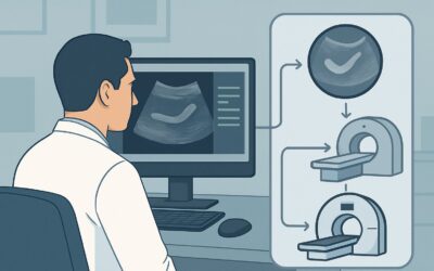
ACR-guided imaging workflow for Child. Suspected acute appendicitis, equivocal or nondiagnostic right lower quadrant ultrasound. Next imaging study. (Suspe

ACR-guided imaging workflow for Child. Suspected acute appendicitis, high clinical risk. Initial imaging. (Suspected Appendicitis-Child): recommended study

ACR-guided imaging workflow for Child. Suspected acute appendicitis, intermediate clinical risk. Initial imaging. (Suspected Appendicitis-Child): recommend

ACR-guided imaging workflow for Child. Suspected acute appendicitis, low clinical risk. Initial imaging. (Suspected Appendicitis-Child): recommended study,

ACR-guided imaging workflow for Adolescent (10 to 17 years of age). Adolescent idiopathic scoliosis. Risk factors. Initial imaging. (Scoliosis-Child): reco

ACR-guided imaging workflow for Adolescent (10 to 17 years of age). Adolescent idiopathic scoliosis. No risk factors. Initial imaging. (Scoliosis-Child): r

ACR-guided imaging workflow for Child (0 to 9 years of age). Early onset idiopathic scoliosis. Initial imaging. (Scoliosis-Child): recommended study, diffe