What Imaging Is Best for Acute Vertigo with Central Neurologic Signs?

It’s 2 a.m. in the emergency department, and you are evaluating a 65-year-old patient with three hours of severe, persistent vertigo, nausea, and gait instability. They are unable to walk without assistance. A standard neurologic exam reveals subtle dysmetria on finger-to-nose testing, and the HINTS (Head-Impulse, Nystagmus, Test-of-Skew) examination is concerning for a central process. You suspect a posterior circulation stroke, but you need to confirm the diagnosis and rule out other dangerous causes. The immediate question is which imaging study to order first.
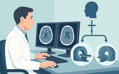
This clinical workflow guide focuses on this specific scenario: an adult patient with acute, persistent vertigo and an abnormal neurologic or HINTS examination suggesting a central etiology. For this presentation, the American College of Radiology (ACR) Appropriateness Criteria rate MRI head without IV contrast as Usually Appropriate, making it the recommended initial imaging study.
Who Fits This Clinical Scenario?
This guidance applies to a well-defined patient population where the pre-test probability of a central nervous system cause of vertigo is high. The key inclusion criteria are:
- Adult patient
- Acute, persistent vertigo (lasting hours to days, not brief, fleeting episodes)
- Abnormal neurologic examination (e.g., new diplopia, dysarthria, limb ataxia, cranial nerve palsies) OR a HINTS examination consistent with a central cause (e.g., a normal head-impulse test in the presence of vertigo, direction-changing nystagmus, or skew deviation).
It is crucial to distinguish this scenario from similar presentations that follow different diagnostic pathways:
- Patient with a normal exam: If the patient has acute persistent vertigo but a completely normal neurologic exam AND a HINTS exam consistent with a peripheral cause (e.g., vestibular neuritis), immediate imaging is often not indicated. This falls under a different ACR variant.
- Patient with triggered, episodic vertigo: If the vertigo is brief (seconds to minutes) and reliably triggered by specific head movements, the likely diagnosis is benign paroxysmal positional vertigo (BPPV). This presentation typically does not require initial imaging.
- Patient with chronic vertigo and hearing loss: If vertigo is chronic or recurrent and associated with unilateral hearing loss or tinnitus, the workup shifts to evaluating the internal auditory canals and cerebellopontine angle, often requiring different MRI protocols.
What Diagnoses Are You Working Up in This Scenario?
When a patient presents with acute vertigo and central signs, the imaging workup is focused on identifying life-threatening conditions affecting the cerebellum and brainstem. The differential diagnosis is narrow and high-stakes.
Posterior Circulation Ischemic Stroke
This is the most critical and common diagnosis to confirm or exclude. Strokes involving the cerebellum or brainstem, supplied by the vertebrobasilar arterial system, classically present with vertigo, ataxia, and other focal deficits. An abnormal HINTS exam can be more sensitive than early MRI for posterior circulation stroke, but imaging is required for confirmation, localization, and to guide therapy.
Cerebellar or Brainstem Hemorrhage
While less common than ischemic stroke, an acute hemorrhage in the posterior fossa is a neurosurgical emergency. Symptoms can be indistinguishable from an ischemic stroke, but the management is drastically different. Rapid identification is paramount to prevent herniation and death.
Demyelinating Disease
In a younger adult, this presentation could represent the initial manifestation of multiple sclerosis (MS). A demyelinating plaque in the brainstem or cerebellum can mimic stroke symptoms. Imaging is key to identifying characteristic lesions and guiding further neurologic workup.
Posterior Fossa Mass
A primary or metastatic tumor, or less commonly an abscess, can present with acute ataxia and vertigo due to mass effect or edema. While often associated with a more subacute onset, acute symptoms can occur due to hemorrhage into the tumor or acute hydrocephalus.
Why Is MRI Head without IV Contrast the Recommended Study?
The ACR designates MRI head without IV contrast as Usually Appropriate because it provides the highest diagnostic yield for the most likely and most dangerous pathologies in this scenario, doing so without radiation or the risks of intravenous contrast.
The key advantage of MRI is its superior soft-tissue resolution and its sensitivity for acute ischemia. Specifically, the Diffusion-Weighted Imaging (DWI) sequence can detect cytotoxic edema from an ischemic stroke within minutes of onset. This is particularly vital in the posterior fossa, an area where CT imaging is often limited by beam-hardening artifact from the dense petrous bones, obscuring the view of the brainstem and cerebellum.
Let’s compare this to other imaging options rated by the ACR for this specific clinical question:
- CT head without IV contrast is rated May be appropriate. Its primary strength is speed and wide availability, making it excellent for rapidly ruling out acute hemorrhage. However, it is notoriously insensitive for acute ischemic stroke, especially in the posterior fossa, where a “normal” early CT can provide false reassurance. It is a reasonable first test only if MRI is immediately unavailable or contraindicated, but a negative result does not exclude an ischemic stroke. This study involves a moderate radiation dose (☢☢☢ 1-10 mSv).
- CTA head and neck with IV contrast is rated May be appropriate (Disagreement). This study is excellent for evaluating the vasculature and can identify vertebral or basilar artery dissection or occlusion. However, it provides less information about the brain parenchyma than a non-contrast MRI and requires both radiation and IV contrast. It is often used as a secondary, problem-solving study after an initial MRI or CT raises suspicion for a vascular etiology.
- MRI head with IV contrast is rated Usually not appropriate for the initial workup. In the hyperacute setting of suspected stroke, gadolinium contrast adds little diagnostic value for identifying ischemia and is not necessary to see hemorrhage. Contrast is typically reserved for follow-up imaging or if the initial non-contrast study suggests a possible tumor, abscess, or demyelinating plaque.
In summary, a non-contrast brain MRI directly visualizes the brain parenchyma to answer the most urgent question: is there an acute stroke, hemorrhage, or mass? It achieves this with the highest sensitivity and no radiation exposure (O 0 mSv), making it the definitive first-line study.
What’s Next After MRI Head without IV Contrast? Downstream Workflow
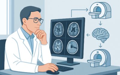
The results of the initial MRI will dictate the subsequent clinical pathway. The workflow branches based on whether the findings are positive, negative, or indeterminate.
If the MRI is positive for acute ischemic stroke:
This is a medical emergency. The immediate next step is an urgent neurology and/or stroke team consultation. Depending on the time from symptom onset and specific findings, the patient may be a candidate for thrombolysis or mechanical thrombectomy. Further vascular imaging with MRA or CTA is almost always required to identify the causative vessel occlusion or stenosis and plan for intervention.
If the MRI is positive for hemorrhage:
This requires an immediate neurosurgical consultation. Management will focus on blood pressure control, reversal of any anticoagulation, and assessing the need for surgical evacuation or external ventricular drain placement, especially if there are signs of hydrocephalus or brainstem compression.
If the MRI is negative:
A negative high-quality MRI, including DWI sequences, makes a significant ischemic stroke highly unlikely. The focus should shift back to a thorough neurologic and otologic evaluation. Consider less common central causes that may not be apparent on initial MRI or reconsider peripheral vestibular causes that can rarely mimic central signs. If clinical suspicion for a vascular event remains very high despite a negative MRI (a rare occurrence), a 24- to 48-hour repeat MRI or vascular imaging may be considered. This may also be the point to consider the sibling scenario of chronic disequilibrium with signs of cerebellar ataxia if symptoms persist.
If the MRI is indeterminate (e.g., shows a non-specific white matter lesion):
Consultation with neurology is recommended. The next step may involve a follow-up MRI with IV contrast to better characterize the lesion, looking for enhancement that might suggest demyelination or a tumor.
Pitfalls to Avoid (and When to Get Help)
In this high-acuity scenario, several common pitfalls can delay diagnosis or lead to misinterpretation.
- Accepting a negative non-contrast CT: The most dangerous pitfall is stopping the workup after a negative non-contrast head CT. Remember, CT is insensitive to early posterior fossa ischemia. If your clinical suspicion for a central cause is high, you must proceed to MRI.
- Delaying the MRI: Time is brain. For a patient with suspected posterior circulation stroke, imaging should be obtained as emergently as possible to facilitate time-sensitive treatments.
- Omitting the DWI sequence: When ordering the MRI, ensure the protocol includes Diffusion-Weighted Imaging. This is the most critical sequence for detecting acute stroke and should never be omitted in this context.
- Misinterpreting the HINTS exam: The HINTS exam is a powerful tool, but it requires proper training to perform and interpret correctly. If you are uncertain, err on the side of caution and obtain imaging.
If the patient shows any signs of declining mental status, worsening neurologic deficits, or severe headache, escalate immediately to your neurology and neurosurgery colleagues, as this may signal brainstem compression or evolving hydrocephalus.
Related ACR Topics and Tools
Navigating imaging guidelines requires access to the right resources. For a comprehensive overview of all clinical variants related to dizziness and ataxia, consult our parent topic hub article. For other tools to help refine your imaging orders, see the links below.
- For breadth across all scenarios in Dizziness and Ataxia, see our parent guide: Dizziness and Ataxia: ACR Appropriateness Decoded.
- To explore other clinical scenarios, use the ACR Appropriateness Criteria Lookup.
- For technical details on the recommended study, browse the Imaging Protocol Library.
- To discuss radiation exposure with patients, use the Radiation Dose Calculator.
Frequently Asked Questions
Why is a non-contrast MRI preferred over an MRI with contrast for this initial workup?
In the acute setting of suspected stroke or hemorrhage, intravenous gadolinium contrast adds little to no diagnostic information and is rated ‘Usually not appropriate’ by the ACR. Acute ischemia is best seen on Diffusion-Weighted Imaging (DWI), and acute hemorrhage is clearly visible on sequences like Gradient Echo (GRE) or Susceptibility-Weighted Imaging (SWI), none of which require contrast. Contrast is reserved for later if there is suspicion of a tumor, abscess, or demyelination.
If my hospital’s MRI scanner is down or the patient has a contraindication, is a CT angiogram (CTA) a good substitute?
A CTA head and neck is rated ‘May be appropriate (Disagreement)’. While it provides excellent detail of the blood vessels and can identify a dissection or occlusion, it is less sensitive than MRI for detecting the resulting brain tissue injury (the stroke itself). It also involves radiation and IV contrast. A non-contrast CT to rule out hemorrhage followed by a CTA is a common pathway when MRI is unavailable, but it is not a direct substitute for the parenchymal detail provided by MRI.
How soon after symptom onset can an MRI detect a posterior circulation stroke?
The Diffusion-Weighted Imaging (DWI) sequence on an MRI is incredibly sensitive and can detect the cellular changes of an ischemic stroke within minutes of onset. This is a major reason why MRI is the preferred modality, as other imaging studies like CT may not show evidence of a stroke for several hours or even days, particularly in the posterior fossa.
What specific findings on a HINTS exam should prompt me to order an MRI?
A HINTS exam is considered ‘central’ or dangerous if it includes any one of the following: a normal Head-Impulse test (no corrective saccade), Nystagmus that is direction-changing in different gazes (or purely vertical/torsional), or a positive Test-of-Skew (vertical misalignment of the eyes). The presence of any of these findings in a patient with acute persistent vertigo is highly suspicious for a brainstem or cerebellar stroke and mandates an urgent MRI.
Does this guidance apply to children with acute vertigo and ataxia?
This specific ACR variant is for adults. While the underlying principles are similar, the differential diagnosis and imaging protocols can differ in children. The ACR provides separate pediatric guidelines, and consultation with a pediatric neurologist or radiologist is recommended when evaluating a child with these symptoms.
Reviewed by Pouyan Golshani, MD, Interventional Radiologist — May 30, 2026