What Imaging Is Best for Routine Asymptomatic Hip Replacement Follow-Up?

A 72-year-old man presents for his scheduled two-year follow-up after a left total hip arthroplasty. He reports no pain, instability, or functional limitations; he has successfully returned to golfing and walking his dog daily. On physical exam, his gait is stable and his hip has an excellent range of motion without discomfort. The orthopedic surgeon now faces a common clinical question: Is routine imaging necessary to screen for silent complications in this well-functioning joint replacement, and if so, which study is the right one to order?
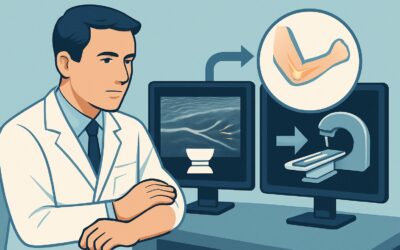
This article provides a focused clinical workflow for this exact scenario, guided by the American College of Radiology (ACR) Appropriateness Criteria. For the routine follow-up of an asymptomatic patient after hip arthroplasty, the ACR designates Radiography hip as Usually Appropriate, establishing it as the standard of care for surveillance.
Who Fits This Clinical Scenario for Asymptomatic Hip Arthroplasty Follow-Up?
This guidance applies specifically to patients who have undergone total hip arthroplasty (THA) and are presenting for a scheduled, routine follow-up visit without any new or worsening symptoms. The key inclusion criterion is the complete absence of clinical complaints related to the prosthetic hip. This includes no pain, clicking, popping, grinding, instability, weakness, or unexplained functional decline. The patient feels the hip is functioning well.
It is critical to distinguish this asymptomatic surveillance scenario from others that require a different diagnostic approach. This workflow does not apply if the patient presents with:
- New or Worsening Symptoms: If the patient reports pain, a sensation of instability, or mechanical symptoms, they fit the “Symptomatic patient with hip prosthesis. Initial imaging” scenario, which has a distinct workup.
- A Recent Fall or Injury: A patient with a history of acute trauma, even if symptoms are mild, should be evaluated under the “Symptomatic hip arthroplasty patient, history of acute injury” criteria, as the concern for periprosthetic fracture is high.
- Signs of Infection: The presence of fever, chills, or local signs like erythema, warmth, or drainage immediately shifts the workup to the “Symptomatic hip arthroplasty patient, infection not excluded” pathway, which often involves laboratory tests and may require more advanced imaging.
- Pain Over the Greater Trochanter: If the pain is localized laterally, suggesting a soft-tissue cause, the “Hip arthroplasty patient with trochanteric pain” scenario is more appropriate.
Correctly identifying the patient as truly asymptomatic is the crucial first step to applying this surveillance imaging strategy.
What Are You Monitoring For in an Asymptomatic Hip Replacement?
In an asymptomatic patient, imaging is not performed to diagnose a cause of pain but rather to conduct surveillance for long-term, silent complications that can progress insidiously. The goal is to identify potential problems before they lead to symptoms or catastrophic component failure. The primary conditions being monitored for are:
Aseptic Loosening
This is the most common reason for long-term failure of a total hip replacement. It is a non-infectious process where the bond between the implant and the bone (or bone cement) weakens over time. On serial radiographs, this can manifest as the development or progression of radiolucent lines at the bone-implant interface. Detecting these changes early allows for planned revision surgery before significant bone loss occurs.
Polyethylene Wear and Osteolysis
The polyethylene (plastic) liner between the metal or ceramic components gradually wears down, producing microscopic debris. The body’s inflammatory response to this debris can trigger bone resorption, a process called osteolysis. This appears on radiographs as focal, scalloped, lytic lesions around the components. Osteolysis can remain silent for years while progressively weakening the surrounding bone, increasing the risk of periprosthetic fracture. Surveillance radiographs are highly effective at detecting osteolysis when it is still manageable.
Component Migration or Subsidence
Serial radiographs allow for precise comparison of the implant’s position and orientation over time relative to bony landmarks. Any change, such as subsidence (sinking) of the femoral stem or a change in the acetabular cup angle, is a clear indicator of implant instability and loosening. These changes often precede the onset of clinical symptoms.
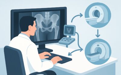
Why Are Hip Radiographs the Recommended Study for Routine Follow-Up?
The ACR designates Radiography hip as Usually Appropriate because it is a low-cost, low-radiation, and highly effective tool for monitoring the key long-term failure modes in an asymptomatic patient. Its value lies in serial comparison.
Standard radiographic views, typically an anteroposterior (AP) pelvis and a frog-leg or true lateral of the prosthetic hip, provide excellent visualization of the bone, the implants, and the critical bone-implant interfaces. This allows for the reliable assessment of component alignment, signs of loosening, polyethylene wear (inferred by component position changes), and osteolysis. The radiation dose is modest (ACR Relative Radiation Level ☢☢☢, 1-10 mSv), which is an important consideration for a test that may be repeated over the patient’s lifetime.
In contrast, more advanced imaging modalities are rated lower for this specific surveillance task:
- MRI hip (without or with contrast): Rated Usually not appropriate. While invaluable for assessing soft tissues in a symptomatic patient, MRI is severely limited by metal susceptibility artifact in the setting of a hip prosthesis. This artifact obscures the bone-implant interface, which is the primary area of interest for surveillance. Specialized metal artifact reduction sequence (MARS) protocols exist but are reserved for specific symptomatic indications, not routine screening.
- CT hip (without contrast): Also rated Usually not appropriate. Though less affected by artifact than MRI, CT still offers little additional information over high-quality radiographs for routine surveillance and imparts a higher radiation dose. Its utility is reserved for complex cases, such as assessing the extent of osteolysis or for pre-operative planning once a problem has already been identified on radiographs.
The clinical consensus is clear: for the asymptomatic patient, the information needed for routine surveillance is best and most efficiently obtained with standard radiographs.
What’s Next After Radiography hip? Downstream Workflow
The results of surveillance radiographs guide the subsequent clinical management and follow-up interval.
- If the radiographs are stable and show no concerns: This is the most common outcome. If there are no signs of loosening, wear, osteolysis, or component migration compared to prior studies, the patient can be reassured. The standard follow-up schedule, as determined by the surgeon (e.g., return in 2-5 years), can be continued. No further imaging is needed at this time.
- If the radiographs are negative but the patient develops symptoms: If the patient becomes symptomatic before their next scheduled visit, the clinical scenario changes. They are no longer an asymptomatic follow-up. The workup should then proceed down the “Symptomatic patient with hip prosthesis” pathway, which may involve laboratory tests (ESR, CRP) and potentially more advanced imaging to investigate the cause of their new symptoms.
- If the radiographs show subtle, non-critical changes: The study may reveal early signs of polyethylene wear or small, stable radiolucent lines. In these cases, the surgeon may opt for “watchful waiting” with a shortened follow-up interval (e.g., return in one year with repeat radiographs) to monitor for progression. This allows for timely intervention if the findings worsen.
- If the radiographs show clear signs of loosening or progressive osteolysis: This finding warrants a more in-depth discussion with the patient about potential revision surgery. Further imaging, such as CT, may be ordered to better characterize the extent of bone loss and to aid in preoperative planning.
Pitfalls to Avoid (and When to Get Help)
When ordering and interpreting surveillance radiographs for a THA, several pitfalls can compromise diagnostic value:
- Inconsistent Radiographic Technique: For serial comparison to be meaningful, imaging technique (e.g., patient positioning, beam angle) must be consistent from one study to the next. Emphasize the need for standardized AP pelvis and true lateral views in the imaging order.
- Not Comparing to Prior Studies: The primary value of surveillance imaging is in detecting change over time. Always review the current radiographs alongside the immediate post-operative films and any other prior studies. A finding may appear significant in isolation but be stable and unchanged for years.
- Overlooking Subtle Signs of Wear: Polyethylene wear is often inferred rather than seen directly. Look for asymmetric positioning of the femoral head within the acetabular cup as a key indicator.
- Misinterpreting Stable Radiolucent Lines: Thin, stable, and non-progressive radiolucent lines (under 2 mm) at the bone-cement interface can represent a stable fibrous membrane rather than loosening. Progression is the key feature of concern.
If radiographs show concerning or equivocal findings of loosening or significant osteolysis, consultation with a musculoskeletal radiologist or an arthroplasty specialist is the appropriate next step.
Related ACR Topics and Tools
For a comprehensive overview of all clinical variants and imaging modalities related to total hip arthroplasty, or to explore the tools used in this workflow, please see the following resources:
- For breadth across all scenarios in Imaging after Total Hip Arthroplasty, see our parent guide: Imaging after Total Hip Arthroplasty: ACR Appropriateness Decoded.
- Imaging Appropriateness Selector — for adjacent scenarios
- Imaging Protocol Library — for technique on the recommended study
- Radiation Dose Calculator — for cumulative dose conversations
Frequently Asked Questions
How often should an asymptomatic patient with a hip replacement get surveillance radiographs?
There is no universal consensus, and practice varies among surgeons. Common protocols involve radiographs at 1, 3, 5, and 10 years post-operatively, and then every 3-5 years thereafter, but this can be adjusted based on patient factors and implant type. The goal is to balance the benefit of early detection against the cost and radiation exposure of imaging.
If a patient has a metal-on-metal hip replacement, is the asymptomatic follow-up different?
Yes. Patients with metal-on-metal (MoM) prostheses are at risk for a specific complication called adverse local tissue reaction (ALTR) from metal ion debris. The ACR has a separate clinical scenario for this situation. Even if asymptomatic, these patients may undergo screening with serum cobalt and chromium levels and potentially cross-sectional imaging like MARS MRI, in addition to radiographs.
Are there any situations where an asymptomatic patient should get an MRI or CT of their hip replacement?
For routine surveillance, no. However, if there is a specific clinical concern despite the lack of symptoms—for example, a known product recall for a specific implant component or a concern for pseudotumor in a MoM hip—then advanced imaging might be considered. These are exceptions and not part of standard asymptomatic follow-up.
Does this guidance apply to hip resurfacing arthroplasty as well?
Yes, the principles of surveillance are similar for hip resurfacing. Radiographs are the primary tool for monitoring component position, loosening, and osteolysis. However, hip resurfacing implants are a form of metal-on-metal articulation, so concerns about metal ion-related complications (ALTR) are also relevant, and follow-up may be more intensive.
What specific radiographic views should be ordered for routine THA follow-up?
A standard surveillance series typically includes an Anteroposterior (AP) view of the pelvis and a true lateral view of the affected hip. The AP pelvis view is crucial for assessing the acetabular component and comparing pelvic landmarks, while the lateral view is essential for evaluating the femoral stem’s position and version.
Reviewed by Pouyan Golshani, MD, Interventional Radiologist — May 26, 2026