What Imaging Should You Order for Chronic Dyspnea with Suspected Pleural or Chest Wall Disease?
A 68-year-old man with a history of construction work presents to your clinic with six months of progressively worsening shortness of breath on exertion. He also describes a persistent, dull ache on his left side. His physical exam is notable for diminished breath sounds at the left lung base and localized tenderness to palpation over the lower ribs. You suspect the cause of his chronic dyspnea originates not from the lung parenchyma itself, but from the pleura or chest wall. You need to select the right initial imaging study to begin the workup. This article details the clinical workflow for this specific scenario, explaining why the American College of Radiology (ACR) finds a standard chest radiograph to be Usually Appropriate as the first-line investigation.
Who Fits This Clinical Scenario?
This guidance applies to adult patients presenting with chronic dyspnea—shortness of breath lasting for weeks to months—where the clinical history and physical examination point toward a pathology of the pleura or chest wall. Key indicators include pleuritic chest pain, localized chest wall tenderness, a history of relevant exposures (like asbestos), prior chest trauma, or physical exam findings such as dullness to percussion or decreased breath sounds suggesting a pleural effusion.
This workflow is specifically for the initial imaging of a suspected pleural or chest wall process. It is crucial to distinguish this from other causes of chronic dyspnea. This guidance does not apply if:
- The etiology is unclear. If there are no localizing signs and the cause of dyspnea is ambiguous, the workup follows a different diagnostic pathway. See the ACR variant for chronic dyspnea of unclear etiology.
- Parenchymal lung disease is the primary suspicion. If the patient has a significant smoking history, cough, and wheezing suggestive of Chronic Obstructive Pulmonary Disease (COPD), the imaging approach is different.
- Diaphragm dysfunction is the specific concern. While related, a primary suspicion of diaphragmatic paralysis or eventration has its own dedicated imaging recommendations, often involving fluoroscopy.
What Diagnoses Are You Working Up in This Scenario?
When you suspect a pleural or chest wall origin for chronic dyspnea, your differential diagnosis is focused on the structures that form the “container” of the lungs. The goal of initial imaging is to detect or rule out these conditions.
Pleural Effusion: This is one of the most common findings. An accumulation of fluid in the pleural space can be transudative (e.g., from heart failure, though this is a cardiovascular origin) or exudative. In this noncardiovascular context, an exudative effusion could be from malignancy (lung cancer, mesothelioma, metastases), infection (parapneumonic effusion or empyema), or inflammatory conditions.
Pleural Thickening or Plaques: Diffuse or localized thickening of the pleura can restrict lung expansion, leading to dyspnea. This is a classic finding in patients with a history of asbestos exposure. Pleural plaques themselves are often asymptomatic but serve as a marker of exposure and may coexist with more serious pathology like mesothelioma.
Chest Wall Tumors: Primary tumors of the chest wall (e.g., sarcomas) or, more commonly, metastatic lesions can cause pain and mechanical restriction. A large mass can compress the lung and limit ventilation, causing a sensation of breathlessness.
Chronic Rib Fractures or Deformity: A history of trauma may lead to non-union of rib fractures or significant chest wall deformity. The resulting pain can cause splinting—shallow breathing to avoid pain—which, over time, can lead to atelectasis and dyspnea.
Why Is a Chest Radiograph the Recommended Initial Study?
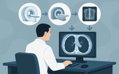
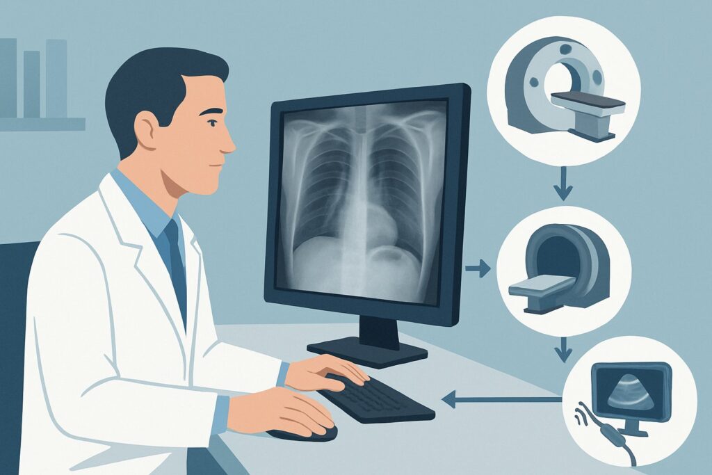
For an initial evaluation of suspected pleural or chest wall disease in a patient with chronic dyspnea, the ACR designates a standard posteroanterior (PA) and lateral Radiography chest as Usually Appropriate. This recommendation is based on an optimal balance of diagnostic utility, accessibility, and safety.
A chest radiograph is highly effective at identifying the most common and significant pathologies on the differential. It can readily detect moderate-to-large pleural effusions, extensive pleural thickening or calcified plaques, obvious rib fractures, and large chest wall masses. Its low radiation dose (Relative Radiation Level ☢ <0.1 mSv) and widespread availability make it the ideal first-line screening tool before committing to more advanced imaging.
Interestingly, both CT chest without IV contrast and CT chest with IV contrast are also rated as Usually Appropriate. This does not mean they should be the first test ordered. Rather, it reflects their role as the definitive problem-solving modality. A chest radiograph is the appropriate initial step, with CT reserved for characterizing an abnormality seen on the radiograph or for cases where clinical suspicion remains high despite a negative or equivocal radiograph.
Other modalities are less suitable for the initial workup:
- US chest is rated Usually Not Appropriate for this indication. While excellent for evaluating a known pleural effusion and guiding thoracentesis, it is a poor screening tool for the entire chest wall and pleura. Its field of view is limited, and it cannot visualize structures deep to bone or aerated lung.
- MRI chest is also Usually Not Appropriate as a first step. MRI offers superb soft-tissue contrast, making it valuable for characterizing a known chest wall mass and its potential invasion into adjacent structures (like the brachial plexus or spine). However, it is more expensive, less available, and more susceptible to motion artifact than CT, making it a secondary, problem-solving tool rather than an initial diagnostic test.
What’s Next After a Chest Radiograph? Downstream Workflow
The results of the initial chest radiograph will guide your next steps. The clinical workflow branches based on whether the study is positive, negative, or indeterminate.
If the radiograph is positive:
- Finding: Pleural Effusion. The next step is often a diagnostic and potentially therapeutic thoracentesis, which can be performed under ultrasound guidance. If malignancy is suspected or the effusion is complex, a CT chest with IV contrast is warranted to evaluate the underlying lung parenchyma and pleura for masses or signs of trapped lung.
- Finding: Pleural Thickening, Plaques, or a Mass. A CT chest is the definitive next step. A non-contrast CT is excellent for assessing calcified plaques, while a contrast-enhanced CT is necessary to characterize soft-tissue masses and assess for enhancement or invasion.
If the radiograph is negative:
A normal chest radiograph significantly lowers the likelihood of a major pleural or chest wall process causing dyspnea. At this point, reconsider the differential diagnosis. Could this be an early interstitial lung disease, small airways disease, or a cardiovascular cause? However, if your clinical suspicion for a pleural or chest wall etiology remains very high based on specific findings (e.g., focal tenderness with a history of cancer), proceeding to a CT chest is a reasonable next step, as it can detect subtle abnormalities missed on radiography.
If the radiograph is indeterminate:
Findings like a blunted costophrenic angle or subtle apical opacity can be equivocal. In these cases, a CT chest is the ideal modality to clarify the finding and determine its significance.
Pitfalls to Avoid (and When to Get Help)
In this clinical scenario, a few common pitfalls can delay diagnosis or lead to unnecessary testing. Be mindful of the following:
- Stopping at a “normal” radiograph: If strong clinical indicators (e.g., a history of asbestos exposure and progressive dyspnea) are present, a normal chest radiograph does not fully exclude pathology. Subtle pleural thickening or an early mesothelioma can be radiographically occult.
- Forgetting the lateral view: A PA-only chest film can miss small posterior pleural effusions or retrosternal masses. Always obtain both PA and lateral views for a complete initial assessment.
- Choosing the wrong CT protocol: If a chest wall mass is suspected, ordering a non-contrast CT may miss important information about vascularity and enhancement. Conversely, for asbestos-related plaques, a non-contrast study is often sufficient.
If a patient presents with acute, severe dyspnea, chest pain, and hemodynamic instability, escalate immediately. This may represent a tension pneumothorax or a massive effusion requiring emergency intervention, not a chronic workup.
Related ACR Topics and Tools
The ACR Appropriateness Criteria are a comprehensive resource for evidence-based imaging decisions. For this scenario and related ones, the following GigHz tools and resources can streamline your workflow and provide deeper context.
- For breadth across all scenarios in Chronic Dyspnea-Noncardiovascular Origin, see our parent guide: Chronic Dyspnea-Noncardiovascular Origin: ACR Appropriateness Decoded.
- To explore imaging guidelines for different clinical presentations, use the ACR Appropriateness Criteria Lookup.
- For detailed procedural techniques, consult the Imaging Protocol Library.
- To discuss radiation exposure with patients, the Radiation Dose Calculator can help quantify and contextualize the dose from recommended studies.
Frequently Asked Questions
Why are both chest radiography and CT chest rated ‘Usually Appropriate’ for this scenario?
The ACR rates studies based on their appropriateness for answering the clinical question, not necessarily their sequence. A chest radiograph is the appropriate *initial* study due to its low cost, low radiation, and high utility for screening. CT is the appropriate *problem-solving* study to characterize findings seen on a radiograph or to investigate further when clinical suspicion is high despite a normal radiograph. Both are appropriate, but they serve different roles in the workflow.
Is a lateral chest radiograph always necessary?
Yes. A two-view (PA and lateral) chest radiograph is the standard of care for initial evaluation. The lateral view is essential for detecting small pleural effusions that only manifest as blunting of the posterior costophrenic angle, evaluating the retrosternal and retrocardiac spaces, and localizing abnormalities seen on the PA view.
What if I suspect a pulmonary embolism (PE) is causing the dyspnea?
This guidance is for chronic dyspnea. A suspected PE is an acute or subacute condition that falls under a completely different diagnostic algorithm. The primary imaging modality for suspected PE is a CT pulmonary angiography (CTPA), which is not addressed in this workflow for chronic, noncardiovascular dyspnea.
When should I consider MRI for a suspected chest wall problem?
MRI is a secondary imaging tool in this context, typically used after a CT has identified a chest wall mass. Its superior soft-tissue contrast makes it the best modality for assessing the extent of a tumor, particularly its potential invasion into the brachial plexus, spinal canal, diaphragm, or adjacent soft tissues.
Does this guidance apply to patients with a history of chest trauma?
Yes, if the presentation is chronic dyspnea. A remote history of trauma could lead to a chronic, non-healing rib fracture, chest wall deformity, or a trapped lung from a remote hemothorax. A chest radiograph is the appropriate first step to look for these sequelae.
Reviewed by Pouyan Golshani, MD, Interventional Radiologist — May 29, 2026