What Is the Best Imaging for a Suspected Foot Foreign Body After Negative X-Rays?

A 45-year-old landscaper presents to your clinic with three months of persistent, localized pain on the plantar aspect of his left foot. He vaguely recalls stepping on something sharp while working but dismissed it at the time. An urgent care visit a month ago included a two-view radiograph of the foot, which was reported as negative for fracture or foreign body. Now, the pain is worsening, and he feels a “small, hard lump” under the skin. You suspect a retained, radiolucent foreign body. The clinical question is clear: with negative initial radiographs, what is the most effective next imaging study to confirm the diagnosis and guide management?
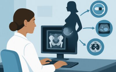
According to the American College of Radiology (ACR) Appropriateness Criteria, for an adult with chronic foot pain and suspected foreign body after negative or indeterminate radiographs, ultrasound (US) of the foot is rated Usually Appropriate. This article provides a detailed workflow for this specific clinical scenario, outlining the differential diagnosis, the rationale for choosing ultrasound over other modalities, and the downstream steps based on the imaging results.
Who Fits This Clinical Scenario for a Suspected Foot Foreign Body?
This guidance is tailored for a specific patient population: an adult presenting with chronic foot pain where the clinical history strongly suggests a retained foreign body. The key inclusion criteria are a history of a puncture wound or penetrating trauma (even if remote or uncertain), localized pain and tenderness, and initial radiographs that are either negative or indeterminate. The chronicity of the pain is also a factor, as it implies a persistent inflammatory response, such as a granuloma, has formed around the object.
It is crucial to distinguish this presentation from similar but distinct clinical scenarios that require a different diagnostic approach. This workflow does not apply if:
- The etiology is unknown. If the patient has diffuse chronic foot pain without a history of trauma or a focal point of tenderness, the workup would fall under the general chronic foot pain scenario, where initial imaging choices may differ.
- A soft-tissue injury is the primary suspicion. If the pain is more consistent with plantar fasciitis, Achilles tendinopathy, or a ligament sprain based on its location and character, the imaging workup should follow the pathway for a suspected tendon or ligament origin.
- An occult fracture is suspected. If the patient has risk factors for a stress fracture (e.g., a runner with new-onset, activity-related pain) and negative radiographs, the next step is often MRI to assess for bone marrow edema, not ultrasound.
Correctly identifying your patient’s scenario ensures the most direct and highest-yield imaging is ordered first.
What Diagnoses Are You Working Up When Radiographs Are Negative?
When a patient presents with a high suspicion for a foreign body despite negative x-rays, your differential diagnosis centers on the object itself and the body’s reaction to it. The goal of imaging is to confirm or exclude these possibilities.
The most common diagnosis you are pursuing is a retained radiolucent foreign body. Many materials that cause puncture wounds are not visible on standard radiographs because they have a similar density to soft tissue. Wood splinters, thorns, plastic fragments, and certain types of glass are notoriously difficult to detect with x-rays. These objects can become encapsulated and lead to chronic, focal inflammation.
A related and often concurrent diagnosis is a foreign body granuloma or abscess. The body’s immune system often walls off a foreign object, creating a firm, painful nodule known as a granuloma. If the object introduced bacteria, a localized abscess can form. In these cases, the patient’s pain is driven by the inflammatory mass or infection, which can be much larger than the foreign body itself. Imaging must be able to characterize this secondary reaction.
Less commonly, the symptoms may be caused by other pathologies that can mimic the sensation of a foreign body. A Morton’s neuroma, a benign thickening of the tissue around a nerve between the metatarsal heads, can cause sharp, localized pain and the feeling of “walking on a pebble.” Similarly, a plantar fibroma, a benign nodule within the plantar fascia, can present as a firm, palpable lump that could be mistaken for an encapsulated foreign object.
Why Is Ultrasound the Recommended Next Step for a Suspected Foreign Body?
For this specific scenario, the ACR designates ultrasound of the foot as Usually Appropriate, making it the clear first-choice modality after inconclusive radiographs. The rationale is based on its high diagnostic yield, safety, and practical advantages.
Ultrasound excels at detecting a wide variety of radiolucent foreign bodies. It uses high-frequency sound waves that can visualize objects like wood, plastic, and glass fragments as hyperechoic (bright) structures, often with a characteristic posterior acoustic shadow. Its sensitivity for detecting these objects is substantially higher than that of plain radiographs. Furthermore, ultrasound can identify secondary signs, such as a surrounding hypoechoic (dark) halo of inflammation, a fluid collection indicating an abscess, or the formation of a granuloma.
A key advantage of ultrasound is its dynamic nature. The sonographer can use the transducer to apply graded pressure directly over the patient’s point of maximal tenderness, correlating the palpable abnormality with the imaging findings in real-time. This ability to localize the pain precisely is invaluable and not possible with static imaging like CT or MRI. This dynamic assessment also allows for evaluation of the object’s relationship to adjacent tendons, nerves, and vessels, which is critical for planning a potential removal. Finally, ultrasound involves no ionizing radiation (0 mSv).
Alternative modalities are rated lower for this initial workup:
- MRI of the foot (without or with contrast) is rated May be appropriate. While excellent for evaluating soft-tissue inflammation, abscesses, and other pathologies like neuromas, it can be less specific in directly identifying the foreign body itself. The object may be obscured by surrounding edema. MRI is more costly, less readily available, and is often better utilized as a problem-solving tool if ultrasound is negative but clinical suspicion remains high.
- CT of the foot (without or with contrast) is also rated May be appropriate. CT is superior to radiographs for detecting materials like glass and gravel but remains limited for organic materials like wood. It also exposes the patient to a small amount of ionizing radiation (adult RRL: ☢ <0.1 mSv). Given the high efficacy of radiation-free ultrasound, CT is not the preferred initial choice.
What’s the Next Step After a Foot Ultrasound?
The results of the foot ultrasound will guide your subsequent management plan, creating a clear decision tree for the patient’s care.
If the ultrasound is positive and clearly identifies a retained foreign body, the next step is typically removal. The ultrasound report should detail the object’s size, location, depth, and proximity to critical structures. This information allows you to refer the patient appropriately to a podiatrist, orthopedic surgeon, or interventional radiologist for excision. For objects that are small or deep, the ultrasound can be used to perform a preoperative skin marking or even provide real-time guidance during the removal procedure to ensure complete extraction and minimize tissue damage.
If the ultrasound is negative and shows no evidence of a foreign body or associated inflammation, you must reconsider the differential diagnosis. If the patient’s symptoms persist, this is an appropriate time to consider an MRI of the foot. The clinical question shifts from “Is there a foreign body?” to “What is the alternative cause of this focal pain?” MRI can effectively diagnose other conditions like a Morton’s neuroma, stress fracture, tendinosis, or plantar fibroma that could be mimicking the initial presentation.
If the ultrasound is indeterminate, for example, showing a focal area of inflammation or a possible granuloma without a clearly defined foreign body within it, MRI can serve as a valuable problem-solving tool. An MRI with intravenous contrast can better characterize the inflammatory process, confirm or rule out an abscess, and may reveal a small, occult foreign body that was obscured by inflammation on the ultrasound.
Common Pitfalls to Avoid in This Workflow
Navigating the workup for a suspected retained foreign body requires careful attention to clinical detail and imaging selection. Avoiding these common pitfalls can prevent diagnostic delays and unnecessary testing.
- Ending the search after negative radiographs. The most significant pitfall is assuming a negative x-ray rules out a foreign body. Many common materials are radiolucent. A persistent, focal point of pain with a plausible history warrants further investigation with ultrasound.
- Submitting a vague imaging requisition. A request for a “foot ultrasound” is insufficient. For a high-yield study, provide the radiologist with a specific history, the suspected material (if known), and clearly indicate the precise location of pain. Marking the patient’s skin with a pen at the point of maximal tenderness before they go for the scan is a highly effective practice.
- Ignoring signs of deep infection. If the ultrasound reveals a large, complex fluid collection suggestive of a deep abscess, or if there is clinical concern for osteomyelitis, the management plan must be escalated. This is no longer a simple foreign body removal and requires urgent surgical consultation for incision, drainage, and debridement.
If the clinical picture is complex or imaging findings are equivocal, consultation with a radiologist or a musculoskeletal specialist is the appropriate next step.
Related ACR Topics and Tools
For a comprehensive overview of imaging for all chronic foot pain scenarios, further reading and specialized tools are available. These resources can help you select the right test for adjacent clinical questions and understand the technical aspects of the recommended studies.
- For breadth across all scenarios in Chronic Foot Pain, see our parent guide: Chronic Foot Pain: ACR Appropriateness Decoded.
- To explore other ACR guidelines or look up different clinical variants, use the ACR Appropriateness Criteria Lookup tool.
- For technical details on how specific studies are performed, consult the Imaging Protocol Library.
- To discuss cumulative radiation exposure with patients, the Radiation Dose Calculator can be a helpful aid.
Frequently Asked Questions
Why not order an MRI immediately if the initial x-ray for a suspected foreign body is negative?
While MRI is an excellent imaging tool for soft tissues, ultrasound is rated higher (*Usually Appropriate*) in this specific scenario for several reasons. Ultrasound is faster, more cost-effective, and has a higher sensitivity for directly visualizing many common radiolucent foreign bodies like wood or plastic. Its dynamic capability, allowing the sonographer to press on the exact point of pain, is a key advantage for localization that MRI lacks. MRI is better reserved as a second-line or problem-solving tool if the ultrasound is negative but clinical suspicion remains high.
What types of foreign bodies are most likely to be missed on a plain radiograph?
Radiographs are excellent for detecting metal and, to a lesser extent, glass and gravel. However, they are very poor at visualizing organic materials, which are common culprits in puncture wounds. Wood splinters, thorns, cactus spines, and plastic fragments are all typically radiolucent, meaning they do not show up on an x-ray. This is why a negative radiograph cannot be used to rule out a retained foreign body.
Can ultrasound be used to guide the removal of the foreign body?
Yes, absolutely. Ultrasound is a powerful tool for both pre-procedural planning and real-time guidance. Before a procedure, a sonographer can use the ultrasound to mark the skin directly over the foreign body. During a procedure, a surgeon or radiologist can use a sterile ultrasound probe to guide their instruments directly to the object, which can help ensure complete removal and minimize damage to surrounding healthy tissue.
If the ultrasound shows inflammation but no clear foreign body, what should I do?
This is an indeterminate finding that requires clinical correlation. The inflammation could be a reaction to a tiny, completely resorbed foreign body, or it could be from another cause like a neuroma or tendinitis. If the patient’s symptoms persist and the diagnosis remains unclear, an MRI is often the best next step. MRI can better characterize the nature of the inflammation and identify other potential soft-tissue pathologies that could be causing the pain.
Does the age of the injury affect the choice of imaging?
For this scenario, the choice of ultrasound as the next step after negative radiographs remains the same regardless of whether the injury is subacute or chronic. However, the chronicity of the injury does change what the ultrasound might show. In an acute injury, you might see the foreign body with some surrounding fluid or hemorrhage. In a chronic case, you are more likely to see a well-defined, hypoechoic granuloma or a thick-walled abscess encapsulating the object.
Reviewed by Pouyan Golshani, MD, Interventional Radiologist — May 30, 2026