What Is the Best Initial Imaging for a Palpable Breast Mass in a Pregnant Patient Under 30?

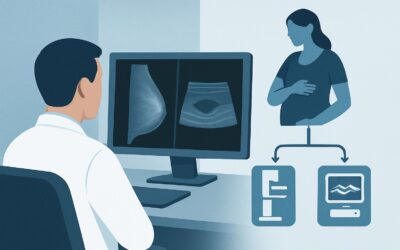
A 28-year-old G1P0 patient at 22 weeks gestation presents to your clinic with a new, palpable lump in her left breast that has been tender for the past two weeks. She is worried, and you recognize the need for a prompt and safe diagnostic workup. The physiological changes of pregnancy, including increased breast density and vascularity, complicate the clinical picture. You need to choose an initial imaging study that is both sensitive for potential pathology and unequivocally safe for the fetus. This article details the American College of Radiology (ACR) evidence-based workflow for this specific scenario, explaining why one modality is the clear first choice. For this patient, the ACR designates breast ultrasound as Usually Appropriate.
Who Fits This Clinical Scenario?
This guidance applies to a very specific patient population: pregnant females under the age of 30 who present with a new focal symptom, such as a palpable mass or focal, non-cyclic pain. The key inclusion criteria are pregnancy, age less than 30, and a localized breast finding that requires initial diagnostic imaging.
It is critical to distinguish this scenario from others that require a different approach. This workflow does not apply to:
- Patients age 30 or older: For pregnant patients aged 30 and above with a palpable mass, the pre-test probability of malignancy is higher, and the imaging recommendations may differ.
- Patients with clinically suspicious nipple discharge: Bloody or spontaneous, unilateral nipple discharge is a distinct clinical problem with its own dedicated ACR Appropriateness Criteria variant.
- Patients requiring breast cancer screening: This guidance is for diagnostic workup of a new symptom, not for routine or high-risk screening during pregnancy, which follows separate protocols.
Applying this workflow to the correct patient presentation ensures an efficient, safe, and evidence-based diagnostic pathway.
What Diagnoses Are You Working Up in This Scenario?
In a young, pregnant patient, the differential diagnosis for a new breast mass is broad, with benign causes being overwhelmingly more common than malignancy. The goal of initial imaging is to characterize the finding and confidently identify or exclude worrisome features.
Galactocele: This is a milk-filled cyst caused by a blocked duct. It is one of the most common benign breast lesions found during pregnancy and lactation. They typically present as smooth, mobile, and sometimes tender masses. Ultrasound is excellent at identifying the classic cystic or complex cystic appearance.
Lactating Adenoma: A benign glandular tumor, this is the most common solid mass to present during pregnancy. It is a variant of a fibroadenoma that undergoes lactational change. While benign, it can grow rapidly due to hormonal stimulation, causing patient anxiety.
Fibroadenoma: These common benign tumors can exist prior to pregnancy and grow in response to hormonal changes, becoming palpable for the first time. They are typically well-circumscribed and mobile.
Abscess or Mastitis: While more common during lactation, focal infection can occur during pregnancy, presenting with pain, erythema, and a tender, palpable mass. Ultrasound can identify a fluid collection that may require drainage.
Pregnancy-Associated Breast Cancer (PABC): Though uncommon in this age group, PABC (defined as breast cancer diagnosed during pregnancy or within one year postpartum) is a critical consideration. Any solid mass that is not clearly benign on imaging requires further evaluation to exclude malignancy. A delay in diagnosis is a primary driver of poorer outcomes in PABC.
Why Is Breast Ultrasound the Recommended Initial Study?
The ACR designates breast ultrasound (US) as Usually Appropriate for this clinical scenario because it directly addresses the diagnostic questions while posing zero risk to the developing fetus. It is the ideal first-line imaging modality for characterizing a palpable abnormality in the dense breast tissue typical of pregnancy.
The primary strengths of ultrasound in this context are:
- Safety: Ultrasound uses sound waves, not ionizing radiation. It has a radiation dose of 0 mSv, making it completely safe for both the mother and the fetus at any stage of gestation.
- High Resolution for Dense Tissue: Pregnancy-related hormonal changes cause glandular tissue to proliferate, significantly increasing mammographic density. This “whited-out” appearance on a mammogram can obscure an underlying mass. Ultrasound is not limited by breast density and provides excellent resolution of focal findings.
- Cystic vs. Solid Differentiation: Ultrasound can definitively determine if a palpable lump is a simple fluid-filled cyst (like a galactocele) or a solid mass, which is the most critical initial step in the workup.
Alternative imaging modalities are rated lower for clear reasons:
- Mammography (Diagnostic): Rated as Usually Not Appropriate. Its sensitivity is significantly reduced by the increased breast density of pregnancy. Furthermore, it involves a low dose of ionizing radiation (ACR RRL: ☢☢ 0.1-1 mSv). While this dose is very low and considered safe for the fetus when the abdomen is shielded, it is an unnecessary exposure when a superior, radiation-free alternative like ultrasound is available.
- MRI Breast Without and With IV Contrast: Rated as Usually Not Appropriate. The primary concern is the use of gadolinium-based contrast agents. Gadolinium is known to cross the placenta and enter the fetal circulation, with unknown long-term consequences. Therefore, its use is avoided during pregnancy unless the potential benefit unequivocally outweighs the potential risk, which is not the case for an initial workup of a palpable mass.
What’s Next After Breast Ultrasound? Downstream Workflow
The results of the breast ultrasound will guide the subsequent steps in the patient’s management. The goal is to provide reassurance for benign findings and to expedite diagnosis for suspicious ones.
If the US shows a simple cyst or a classic galactocele (BI-RADS 2): No further imaging is typically needed. The patient can be reassured of the benign nature of the finding. If the cyst is large and painful, ultrasound-guided aspiration can be performed for symptomatic relief.
If the US shows a probably benign solid mass (BI-RADS 3): For a finding with classic benign features, such as a typical fibroadenoma, the standard recommendation is short-term imaging follow-up. This might involve a repeat ultrasound in 3-6 months, which could be performed during the third trimester or in the early postpartum period to ensure stability.
If the US shows an indeterminate or suspicious solid mass (BI-RADS 4 or 5): Any solid mass that does not meet strict criteria for being benign requires tissue sampling. The next step is an ultrasound-guided core needle biopsy. This procedure is safe to perform during pregnancy, uses local anesthetic, and is essential for obtaining a definitive histologic diagnosis. Delaying biopsy until after delivery is not recommended as it can lead to a significant delay in cancer diagnosis.
If the US is negative but a palpable lump persists: This situation requires close clinical correlation. The radiologist and referring clinician should confer. Depending on the degree of clinical suspicion, options include a short-interval clinical follow-up or, in rare cases, proceeding to diagnostic mammography with abdominal shielding if suspicion remains high.
Pitfalls to Avoid (and When to Get Help)
Navigating a breast concern during pregnancy requires vigilance to avoid common missteps. Be aware of these potential pitfalls:
- Attribution Error: Do not automatically attribute a new, persistent palpable mass to “normal pregnancy changes.” While most are benign, this assumption can delay the diagnosis of PABC.
- Delaying the Workup: Avoid the temptation to “watch and wait” until after delivery for a persistent, palpable solid mass. The diagnostic workup, including ultrasound and biopsy if needed, can and should proceed promptly.
- Ordering the Wrong First Test: Starting with mammography in this young, pregnant population is a common error. It is a lower-yield test due to breast density and involves unnecessary radiation exposure when ultrasound is the superior first choice.
- Hesitancy to Biopsy: A suspicious finding on ultrasound warrants a biopsy. Ultrasound-guided core needle biopsy is the standard of care and is safe during pregnancy.
If an ultrasound reveals a suspicious (BI-RADS 4 or 5) lesion, immediate escalation for a biopsy is the appropriate next step. Consultation with a breast surgeon is recommended.
Related ACR Topics and Tools
This article covers one specific clinical variant. For a comprehensive overview of all scenarios related to breast imaging during pregnancy, or to explore the tools used to create these evidence-based guidelines, please refer to the following resources.
- For breadth across all scenarios in Breast Imaging During Pregnancy, see our parent guide: Breast Imaging During Pregnancy: ACR Appropriateness Decoded.
- To look up other clinical scenarios, use the ACR Appropriateness Criteria Lookup tool.
- To review standardized imaging techniques, see the Imaging Protocol Library.
- To understand and discuss radiation exposure with patients, consult the Radiation Dose Calculator.
Frequently Asked Questions
Is a breast ultrasound really safe for the baby during pregnancy?
Yes, a breast ultrasound is completely safe for both the mother and the fetus. It uses high-frequency sound waves, not ionizing radiation, to create images. There is no known risk to the developing baby at any stage of pregnancy.
Why isn’t mammography the first choice for a palpable lump in a pregnant patient under 30?
There are two main reasons. First, hormonal changes during pregnancy cause breast tissue to become much denser, which can make it very difficult for a mammogram to detect an underlying mass. Second, while the radiation dose from a mammogram is very low and considered safe with abdominal shielding, it’s best to avoid any unnecessary radiation exposure to the mother during pregnancy when a non-radiation alternative like ultrasound is more effective for this specific clinical question.
What happens if the ultrasound is negative, but I can still feel the lump?
This is an important situation that requires close collaboration between you and the radiologist. It’s called clinical-radiologic discordance. The next step is typically a careful repeat clinical breast exam. Depending on the level of suspicion, options may include a short-term follow-up clinical exam in a few weeks or, if concern persists, considering a diagnostic mammogram despite the limitations.
If a biopsy is needed, is it safe to perform during pregnancy?
Yes, an ultrasound-guided core needle biopsy is a safe and standard procedure during pregnancy. It is performed with a local anesthetic (like lidocaine), which has a well-established safety profile. It is the definitive way to diagnose a suspicious solid mass and should not be delayed until after delivery.
If the ultrasound finds a benign issue like a galactocele, does it need to be removed?
Generally, no. A galactocele is a benign, milk-filled cyst. Once diagnosed on ultrasound, it does not require removal unless it is causing significant pain or discomfort. In those cases, it can be drained with a needle (aspirated) under ultrasound guidance for symptomatic relief.
Reviewed by Pouyan Golshani, MD, Interventional Radiologist — May 30, 2026