What Is the Best Initial Imaging for Acute Blunt or Penetrating Hand Trauma?
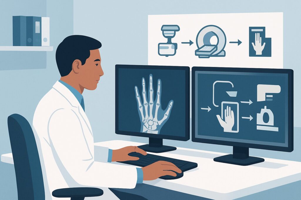
A 34-year-old construction worker presents to the urgent care clinic after a heavy tool fell on their right hand. They report immediate pain, swelling, and difficulty making a fist. On exam, there is focal tenderness over the third metacarpal and diffuse dorsal hand edema. You suspect a fracture, but the exact location and extent are unclear. This is the classic clinical decision point for initial imaging in acute hand trauma. This article provides a detailed workflow for this specific scenario, guiding you through the American College of Radiology (ACR) Appropriateness Criteria. For this presentation, the ACR designates Radiography of the area of interest as Usually appropriate, establishing it as the clear first-line imaging study.
Who Fits This Clinical Scenario?
This guidance applies to adult and pediatric patients presenting with a history of recent, acute trauma to the hand or wrist. The mechanism can be either blunt (e.g., a fall, a crush injury, a direct blow) or penetrating (e.g., from a nail, glass shard, or other sharp object). The key element is that this is the initial imaging workup, performed before any other studies have been obtained.
This workflow is specifically for the undifferentiated first presentation. It is crucial to distinguish this from related but distinct clinical questions. This guidance does not apply if:
- Initial radiographs are negative or equivocal, but clinical suspicion for fracture remains high. This situation, particularly common with suspected scaphoid fractures, constitutes a different clinical scenario requiring consideration of advanced imaging like MRI or CT.
- A fracture is already identified on radiographs, and the primary question is about associated soft tissue injury. If you are concerned about tendon rupture or significant ligamentous disruption in the setting of a known fracture, the imaging workup shifts focus, often toward MRI.
- The primary concern is a retained foreign body after penetrating trauma, and initial radiographs are negative. If you suspect a non-radiopaque foreign body (like wood or plastic), ultrasound is often the next best step, which falls under a separate ACR variant.
What Diagnoses Are You Working Up in This Scenario?
When ordering the initial imaging for acute hand and wrist trauma, the primary goal is to identify or exclude significant osseous and articular injuries that require specific management. The differential diagnosis is focused on conditions readily detectable by radiography.
Fractures are the most common and critical diagnosis to confirm or rule out. This includes fractures of the eight carpal bones (e.g., scaphoid, triquetrum), the five metacarpals (e.g., a Boxer’s fracture of the fifth metacarpal neck), and the fourteen phalanges. Radiographs are highly effective at identifying fracture lines, displacement, angulation, and intra-articular extension, all of which are critical for determining treatment, whether it be splinting, casting, or surgical fixation.
Dislocations and Subluxations are also a key consideration. These injuries involve the complete or partial malalignment of a joint, such as the distal radioulnar joint (DRUJ), the carpometacarpal (CMC) joints, or the interphalangeal joints. A perilunate dislocation, for example, is a severe wrist injury that is a true orthopedic emergency. Plain radiographs are the cornerstone of diagnosis, allowing for rapid assessment of joint congruity.
Radiopaque Foreign Bodies are a primary concern in penetrating trauma. Metallic fragments, glass, gravel, and other dense materials are often clearly visible on standard radiographs. Identifying the presence, location, and size of a foreign body is essential for planning its potential removal and managing the risk of infection or injury to adjacent neurovascular structures.
Why Is Radiography the Recommended Initial Study for This Presentation?
The ACR rates Radiography of the area of interest as Usually appropriate because it directly and efficiently addresses the most common and urgent clinical questions in acute hand and wrist trauma. It provides an excellent balance of diagnostic utility, accessibility, speed, and safety as a first-line investigation.
Standard radiographic series of the hand or wrist are highly sensitive and specific for detecting the vast majority of fractures and dislocations. The technique is fast, widely available in nearly all emergency and outpatient settings, and provides the necessary information to guide immediate management, such as reduction, splinting, and referral to an orthopedic specialist. The radiation dose is variable depending on the specific views obtained but is generally low, making it a safe initial choice for both adult and pediatric patients.
In contrast, more advanced imaging modalities are rated lower for this initial workup:
- CT of the area of interest (without or with contrast) is rated Usually not appropriate. While CT provides exquisite bony detail and is invaluable for characterizing complex, intra-articular fractures, it is not the right first step. It involves a higher radiation dose and cost compared to radiography and is best reserved for pre-operative planning or for clarifying findings that are equivocal on initial plain films.
- MRI of the area of interest (without or with contrast) is also rated Usually not appropriate. MRI is the gold standard for evaluating soft tissues like ligaments, tendons, and cartilage, as well as detecting occult fractures (bone marrow edema). However, it is more expensive, less available, and more time-consuming than radiography. Its use is best reserved for follow-up imaging when initial radiographs are negative but a significant ligamentous injury or occult fracture is still suspected.
- Ultrasound (US) of the area of interest is rated Usually not appropriate for the primary evaluation of a potential fracture. While US can detect cortical disruptions and is excellent for evaluating soft tissues and non-radiopaque foreign bodies, it is highly operator-dependent and is not the standard modality for a comprehensive initial bone survey of the hand or wrist.
What’s Next After Radiography? Downstream Workflow
The results of the initial radiographs will dictate the subsequent clinical pathway. The decision tree is relatively straightforward and is designed to ensure timely and appropriate care.
If the radiograph is POSITIVE for a fracture or dislocation: The next step is definitive management. This typically involves immobilization (splinting or casting), pain control, and a referral to an orthopedic or hand surgeon. For complex injuries, such as those with significant displacement, angulation, or intra-articular involvement, the consulting specialist may order a CT scan to better define the fracture pattern and plan for potential surgical intervention.
If the radiograph is NEGATIVE but clinical suspicion remains high: This is a common and important clinical branch point. A patient with classic “snuffbox” tenderness but negative initial wrist radiographs, for example, should be treated as having a presumptive scaphoid fracture. The next step is typically immobilization in a thumb spica splint and arranging for follow-up imaging. This moves the patient into a different clinical scenario: “Suspect acute hand or wrist trauma. Initial radiographs negative or equivocal. Next imaging study.” The appropriate next study in that case is often an MRI without contrast to look for an occult fracture.
If the radiograph is EQUIVOCAL: Sometimes, findings are unclear due to patient positioning, overlying structures, or subtle abnormalities. In these cases, obtaining additional radiographic views can be helpful. If uncertainty persists, the decision to pursue advanced imaging with CT or MRI depends on the specific anatomical location and the suspected injury.
Pitfalls to Avoid (and When to Get Help)
Several common pitfalls can occur when managing acute hand and wrist trauma. First, failing to order the appropriate, dedicated views for a specific area of concern can lead to a missed diagnosis. For example, if you suspect a scaphoid fracture, a standard wrist series is insufficient; you must specifically order a “scaphoid series.” Second, be wary of dismissing persistent, focal pain after a negative radiograph—this is the classic presentation of an occult fracture. Third, in penetrating trauma, remember that not all foreign bodies (e.g., wood, plastic) are radiopaque; a negative x-ray does not rule out a retained object. Finally, do not let imaging delay the assessment and management of neurovascular compromise or a suspected compartment syndrome, which are clinical diagnoses requiring immediate surgical consultation.
Related ACR Topics and Tools
This article covers one specific scenario in depth. For a broader view of the parent topic or to explore related imaging guidelines and tools, the following resources are available:
- For breadth across all scenarios in Acute Hand and Wrist Trauma, see our parent guide: Acute Hand and Wrist Trauma: ACR Appropriateness Decoded.
- To explore other clinical presentations, use the ACR Appropriateness Criteria Lookup.
- For details on imaging techniques, consult the Imaging Protocol Library.
- To discuss radiation exposure with patients, use the Radiation Dose Calculator.
Frequently Asked Questions
Why not get a CT scan first for a severe-looking hand injury?
While CT provides excellent bone detail, it is rated ‘Usually not appropriate’ as the initial study by the ACR for this scenario. A standard radiograph is faster, uses significantly less radiation, is more widely available, and is sufficient to diagnose the vast majority of fractures and dislocations. CT is best used as a secondary study to better characterize complex fractures identified on radiographs or for pre-operative planning.
What specific radiographic views should I order?
The order should specify the area of interest. A standard ‘hand series’ typically includes posteroanterior (PA), oblique, and lateral views. A ‘wrist series’ includes PA, oblique, and lateral views. If you have a specific clinical concern, order dedicated views—for example, a ‘scaphoid series’ (which includes a PA view with ulnar deviation) is crucial if you suspect a scaphoid fracture.
What if I suspect a scaphoid fracture but the initial x-ray is negative?
This is a critical exception. Due to the risk of avascular necrosis with a missed scaphoid fracture, a negative initial radiograph in the setting of high clinical suspicion (e.g., snuffbox tenderness) is not sufficient to rule out injury. The patient should be immobilized in a thumb spica splint and scheduled for follow-up imaging, typically an MRI without contrast, which is highly sensitive for detecting occult fractures.
Can a radiograph reliably find a foreign body from penetrating trauma?
Radiographs are excellent for detecting radiopaque foreign bodies, such as metal, glass, and gravel. However, they cannot visualize radiolucent materials like wood, thorns, or plastic. If you suspect a retained foreign body of this type and the radiograph is negative, the next appropriate imaging study is typically a high-resolution ultrasound.
Is an ultrasound ever the right first choice in acute hand trauma?
For the initial evaluation of a suspected fracture or dislocation, the ACR deems ultrasound ‘Usually not appropriate.’ Its primary role in acute hand and wrist trauma is for evaluating soft tissue structures (tendons, ligaments), assessing for fluid collections like an abscess, or locating suspected non-radiopaque foreign bodies after initial radiographs are negative.
Reviewed by Pouyan Golshani, MD, Interventional Radiologist — May 29, 2026