What Is the Best Next Imaging Study for Chronic Knee Pain with Degenerative Radiographs?
A 64-year-old patient presents to your clinic with a six-month history of worsening right knee pain. An initial radiograph ordered by their primary care physician demonstrates moderate medial compartment degenerative changes and small osteophytes, consistent with osteoarthritis. However, the patient now reports new mechanical symptoms, including painful catching and a sensation of locking, which are limiting their ability to walk. You have a diagnosis of osteoarthritis from the X-ray, but the clinical picture suggests a superimposed problem. What is the most appropriate next imaging study to evaluate the internal structures of the knee and guide potential intervention?
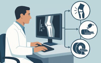
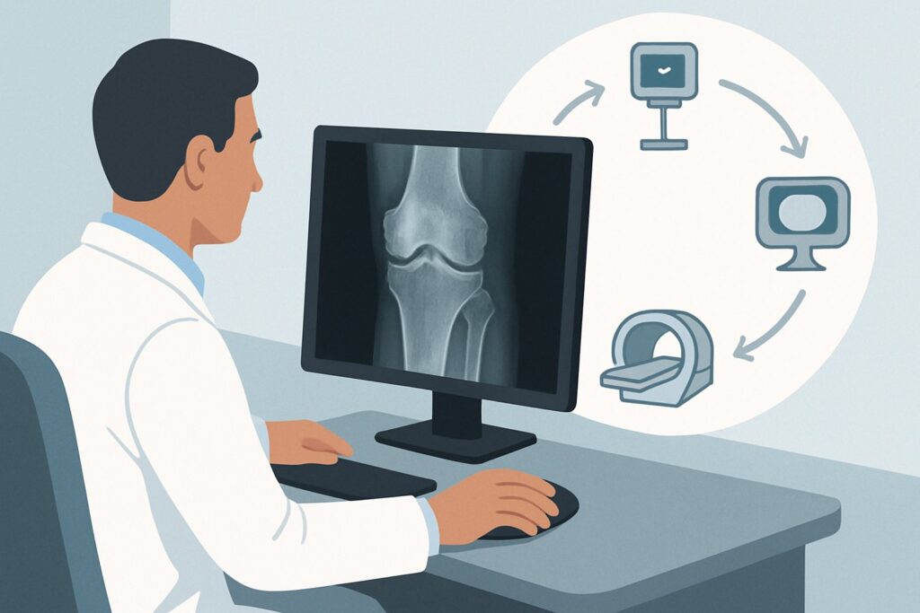
This article provides a focused workflow for this exact clinical question, based on the American College of Radiology (ACR) Appropriateness Criteria. For this scenario, where an initial radiograph shows degenerative changes or chondrocalcinosis, an MRI of the knee without IV contrast is rated as ‘May be appropriate’ to further characterize the source of pain and mechanical symptoms.
Who Fits This Clinical Scenario for Chronic Knee Pain?
This guidance applies to a specific subset of patients: adults or children aged 5 years or older with chronic knee pain where the initial diagnostic step—a knee radiograph—has already been performed and is positive for specific findings. The key inclusion criteria are:
- Chronic Knee Pain: The symptoms are persistent or recurrent, not from an acute, high-impact traumatic event.
- Prior Radiograph Performed: The patient is not presenting for their first imaging study.
- Specific Radiographic Findings: The X-ray must show evidence of either degenerative changes (e.g., joint space narrowing, osteophytes, subchondral sclerosis) or chondrocalcinosis.
It is critical to distinguish this situation from similar but distinct clinical presentations that follow different diagnostic pathways. This workflow does not apply if:
- No initial imaging has been performed. That patient falls under the initial imaging scenario for chronic knee pain.
- The initial knee radiograph was negative. A negative X-ray requires a different workup, as the differential diagnosis shifts away from complications of known osteoarthritis.
- The radiograph shows a clear osteochondral lesion or signs of inflammatory arthritis. These findings point toward more specific ACR scenarios with tailored imaging recommendations.
This pathway is for when the radiograph confirms a chronic process like osteoarthritis, but the severity of symptoms or new mechanical features suggest an internal derangement that the X-ray cannot visualize.
What Diagnoses Are You Working Up When Radiographs Already Show Degeneration?
When a radiograph confirms osteoarthritis or chondrocalcinosis, the goal of subsequent imaging is not to re-diagnose the degenerative process but to identify superimposed, treatable pathologies that explain the patient’s current symptoms. The differential is focused on soft tissue and bone marrow abnormalities that are occult on X-ray.
Meniscal Tear: This is a primary consideration, especially with new mechanical symptoms like locking, catching, or giving way. Degenerative meniscal tears are common in the setting of osteoarthritis and can be a significant pain generator. MRI is the gold standard for visualizing meniscal morphology and identifying tears that may be amenable to arthroscopic intervention.
Articular Cartilage Injury: Radiographs infer cartilage loss through joint space narrowing, but they cannot directly visualize the cartilage surface. A focal, full-thickness chondral defect or delamination can cause significant pain and mechanical symptoms disproportionate to the overall degree of arthritis. MRI provides detailed evaluation of cartilage integrity.
Subchondral Insufficiency Fracture or Osteonecrosis: These conditions can present as acute-on-chronic pain in an osteoarthritic knee. They represent stress failure of the subchondral bone or localized bone death, respectively. Early radiographic findings are often absent or subtle, but MRI is highly sensitive for the characteristic bone marrow edema patterns associated with these diagnoses.
Intra-articular Loose Body: While large, ossified loose bodies may be visible on radiographs, smaller cartilaginous fragments are not. These can cause intermittent locking and pain. MRI can identify both osseous and non-osseous loose bodies within the joint.
Why Is MRI Without Contrast the Next Step for Knee Pain with Degenerative Radiographs?
In this scenario, the ACR rates MRI of the knee without IV contrast as ‘May be appropriate’. This rating reflects that while not every patient with osteoarthritis on X-ray needs an MRI, it becomes a reasonable and often necessary next step when symptoms are severe, mechanical, or discordant with the radiographic findings. The rationale is based on MRI’s superior soft-tissue and bone marrow contrast, which directly addresses the key differential diagnoses.
MRI excels at visualizing the menisci, ligaments, articular cartilage, and subchondral bone—the very structures that X-rays cannot assess. It can definitively identify a meniscal tear, characterize the size and depth of a chondral defect, or detect a subchondral insufficiency fracture, providing crucial information for surgical planning or conservative management.
Alternative studies are rated lower for specific reasons in this context:
- CT knee without IV contrast is also rated ‘May be appropriate’ but is generally less preferred. While excellent for bone detail and detecting osseous loose bodies, it provides significantly less information about the menisci, cartilage, and bone marrow compared to MRI. Its use of ionizing radiation (ACR RRL: ☢ <0.1 mSv for adults) also makes it less ideal when a radiation-free option exists.
- Ultrasound (US) of the knee is rated ‘Usually not appropriate’. While useful for evaluating superficial structures, effusions, and tendons, it cannot adequately assess the deep structures of the knee, such as the cruciate ligaments, menisci, or subchondral bone, which are the primary targets of this workup.
An MRI without intravenous contrast is sufficient for this indication. The addition of contrast (rated ‘Usually not appropriate’) is generally unnecessary for evaluating the degenerative and traumatic pathologies in this differential and should be reserved for specific indications like suspected tumor, infection, or inflammatory synovitis. The lack of ionizing radiation (ACR RRL: O 0 mSv) makes MRI a safe choice for both adult and pediatric patients.
Once you’ve decided on an MRI of the knee without contrast, our protocol guide covers the essential sequences, patient positioning, and interpretation principles. MRI Knee Without Contrast.
What’s Next After MRI Without Contrast? Downstream Workflow
The results of the knee MRI will guide the subsequent clinical management, which can range from continued conservative therapy to surgical consultation. The downstream workflow depends directly on the findings.
- If the MRI confirms a significant meniscal tear or large chondral defect: A referral to an orthopedic surgeon is the appropriate next step. The MRI findings will be essential for determining if the patient is a candidate for arthroscopy, debridement, repair, or other cartilage restoration procedures. The presence of severe background osteoarthritis may influence the surgical approach.
- If the MRI shows a subchondral insufficiency fracture: Management is typically conservative, involving a period of protected or non-weight-bearing to allow the bone to heal. The MRI confirms the diagnosis and rules out other causes, preventing unnecessary interventions.
- If the MRI shows only degenerative changes consistent with the radiograph and no new pathology: This negative result is also clinically valuable. It confirms that the patient’s symptoms are likely due to the progression of their osteoarthritis. The focus should be on optimizing non-operative management, including physical therapy, intra-articular injections, bracing, and analgesia.
- If the MRI is indeterminate or suggests an unexpected finding (e.g., synovitis suggesting inflammatory arthropathy): Further workup may be needed. This could involve rheumatologic consultation and laboratory testing or, in rare cases, a follow-up MRI with contrast or image-guided aspiration.
Pitfalls to Avoid (and When to Get Help)
Navigating this clinical scenario requires avoiding several common pitfalls to ensure timely and appropriate care.
- Pitfall 1: Prematurely ordering an MRI. Do not skip the initial weight-bearing radiographs. They are essential for assessing joint space narrowing and alignment, which are critical for long-term management and surgical planning.
- Pitfall 2: Ordering the wrong MRI protocol. An MRI with IV contrast is ‘Usually not appropriate’ for this indication and adds unnecessary cost and potential risk. Specify “MRI knee without IV contrast” to evaluate for internal derangement.
- Pitfall 3: Over-interpreting MRI findings. In an older population with osteoarthritis, incidental findings like degenerative meniscal signal are common and may not be the source of pain. Correlate MRI findings directly with the patient’s specific symptoms and physical exam.
- Pitfall 4: Ignoring referred pain. Chronic knee pain, especially in older adults, can be referred from the hip. If the knee MRI is unrevealing but symptoms persist, consider a clinical and radiographic evaluation of the ipsilateral hip.
If the clinical picture and imaging findings are complex or discordant, consultation with a musculoskeletal radiologist or an orthopedic surgeon can provide valuable guidance on the next steps.
Related ACR Topics and Tools
This article covers one specific scenario within the broader topic of chronic knee pain. The ACR provides guidance on numerous related presentations, and understanding the appropriate study for each is key to efficient and effective care. For breadth across all scenarios in Chronic Knee Pain, see our parent guide: Chronic Knee Pain: ACR Appropriateness Decoded.
For additional decision support and technical details, the following GigHz resources are available:
- ACR Appropriateness Criteria Lookup — for adjacent scenarios not covered here.
- Imaging Protocol Library — for technique on the recommended study.
- Radiation Dose Calculator — for cumulative dose conversations with patients.
Frequently Asked Questions
Why is MRI only ‘May be appropriate’ and not ‘Usually appropriate’ if it’s the best next test?
The ‘May be appropriate’ rating reflects clinical nuance. Not every patient with degenerative changes on an X-ray needs an MRI. The progression to MRI is appropriate only when the clinical symptoms (like locking, instability, or pain disproportionate to radiographic findings) suggest a superimposed internal derangement. If the symptoms are consistent with the degree of osteoarthritis seen on the radiograph, an MRI may not change management and is therefore not automatically indicated.
Is a CT scan a reasonable alternative to MRI in this scenario?
According to the ACR, a CT knee without IV contrast is also rated ‘May be appropriate’. However, it is generally considered a secondary option. While CT is excellent for evaluating complex fractures and osseous loose bodies, it provides poor visualization of the menisci, ligaments, and cartilage. Since these soft-tissue structures are the primary targets of the workup, MRI is almost always preferred. CT may be considered if the patient has a contraindication to MRI.
Should I order an MRI with contrast if I suspect something more than just a meniscal tear?
For this specific scenario—evaluating for internal derangement superimposed on osteoarthritis—IV contrast is ‘Usually not appropriate’. A non-contrast MRI provides excellent detail for menisci, cartilage, and subchondral bone. Contrast is typically reserved for cases where there is a specific concern for infection, inflammatory arthritis (synovitis), or a tumor, which fall under different clinical scenarios.
What if the patient has chondrocalcinosis on the radiograph but no degenerative changes?
This workflow still applies. The presence of chondrocalcinosis (cartilage calcification, often associated with CPPD) on a radiograph places the patient in this same ACR variant. An MRI without contrast would still be rated ‘May be appropriate’ to evaluate for underlying meniscal or chondral pathology that could be causing the patient’s chronic pain.
Does this guidance apply to a patient with a known history of osteoarthritis who now has an acute injury?
No, this guidance is for chronic knee pain. If a patient with pre-existing osteoarthritis sustains a new, acute traumatic injury (e.g., a fall with a twisting motion), the workup would follow the ACR guidelines for Acute Knee Trauma, which is a separate topic with different recommendations.
Reviewed by Pouyan Golshani, MD, Interventional Radiologist — May 29, 2026