What Is the Best Supplemental Breast Screening for Average-Risk, Heterogeneously Dense Breasts?

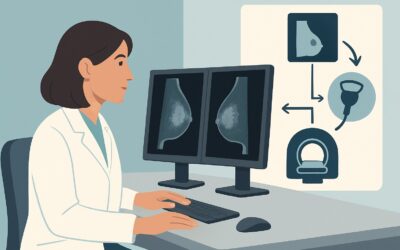
A 48-year-old woman with no significant personal or family history of breast cancer is in your office for her annual wellness visit. Her screening mammogram report from last week is on your screen, noting BI-RADS C “heterogeneously dense” breast tissue. The patient is asking if she needs any additional testing, having read online that dense breasts can “hide” cancers. You need to decide on the appropriate supplemental screening strategy for this common clinical scenario. This article outlines the evidence-based workflow for an average-risk woman with heterogeneously dense breasts, a situation where the American College of Radiology (ACR) has specific guidance. For this patient, the ACR deems Digital Breast Tomosynthesis (DBT) screening as Usually Appropriate.
Who Fits This Clinical Scenario?
This guidance applies to a specific patient population: adult females undergoing supplemental breast cancer screening who are at average risk and have heterogeneously dense breasts. It is critical to confirm both criteria before proceeding.
Inclusion Criteria:
- Average Risk: This category includes women with less than a 15% lifetime risk of breast cancer. They have no personal history of breast cancer, ductal carcinoma in situ (DCIS), or lobular carcinoma in situ (LCIS). They also lack a known high-risk genetic mutation (e.g., BRCA1/2) and have not undergone chest radiation therapy before age 30. A strong family history (e.g., a first-degree relative with premenopausal breast cancer) would move a patient into an intermediate or high-risk category.
- Heterogeneously Dense Breasts: This corresponds to Breast Imaging Reporting and Data System (BI-RADS) density category C. On a mammogram, this means there are scattered areas of fibroglandular density, which could obscure small masses. This category accounts for approximately 40% of the screening population.
Exclusion Criteria: This workflow is not appropriate for patients who fall into other risk or density categories. For example, a patient with extremely dense breasts (BI-RADS category D) or a patient with a known BRCA1 mutation (high risk) would follow a different screening algorithm, often involving breast MRI. Similarly, a woman with nondense (fatty or scattered fibroglandular) breasts does not require supplemental screening beyond standard mammography.
What Diagnoses Are You Working Up in This Scenario?
In a screening context, the goal is not to work up a symptom but to detect occult disease before it becomes clinically apparent. The primary concern in a patient with heterogeneously dense breasts is the potential for “masking,” where the dense fibroglandular tissue obscures an underlying malignancy on a standard two-dimensional (2D) mammogram.
Invasive Ductal Carcinoma (IDC): This is the most common type of invasive breast cancer. In dense breasts, a small, spiculated IDC can be difficult to distinguish from normal overlapping glandular tissue on a 2D mammogram. Supplemental screening aims to unmask these early, treatable cancers.
Ductal Carcinoma In Situ (DCIS): As a non-invasive, precursor lesion, DCIS often presents as microcalcifications but can also form a non-calcified mass. Like IDC, a non-calcified DCIS can be hidden by dense tissue, and supplemental imaging can improve its detection.
Invasive Lobular Carcinoma (ILC): This less common but consequential subtype is notoriously difficult to detect mammographically. ILC often grows in a diffuse, single-file pattern without forming a discrete mass, making it particularly susceptible to masking by dense breast tissue. Improved imaging techniques are crucial for its early detection.
Summation Artifacts (False Positives): A key “diagnosis” to rule out is a false positive caused by overlapping normal tissue. On a 2D image, benign glands can superimpose to create the appearance of a suspicious mass (a summation artifact). A primary goal of supplemental screening is to reduce these artifacts, thereby decreasing the patient recall rate and the associated anxiety and cost.
Why Is Digital Breast Tomosynthesis Screening the Recommended Study for This Presentation?
For an average-risk woman with heterogeneously dense breasts, the ACR Appropriateness Criteria rate Digital Breast Tomosynthesis (DBT) screening as Usually Appropriate. DBT, often referred to as 3D mammography, is the preferred initial study because it directly addresses the primary diagnostic challenge in this scenario: the masking effect of overlapping fibroglandular tissue.
DBT acquires multiple low-dose X-ray images of the breast from various angles during a short sweep. A computer then reconstructs these images into a series of thin, one-millimeter slices. By allowing the radiologist to scroll through the breast tissue layer by layer, DBT effectively “unpeels” the overlapping structures that can hide a cancer on a flat 2D image. This technique has consistently been shown to increase the cancer detection rate while simultaneously decreasing the recall rate for non-cancerous findings.
The radiation dose for a combined 2D mammogram and DBT is low (ACR Relative Radiation Level ☢☢, 0.1-1 mSv), well within accepted safety limits for annual screening and only slightly higher than 2D mammography alone. Many modern systems can now generate a synthetic 2D image from the DBT data, avoiding the extra radiation dose of a separate 2D acquisition.
Why are other studies rated lower?
- Breast Ultrasound (US): While rated as May be appropriate, handheld screening ultrasound has significant limitations. It is highly operator-dependent, has a lower specificity than DBT, and leads to a substantially higher rate of false-positive findings and benign biopsies. While it can detect some cancers missed by mammography, its high false-positive rate makes DBT a better initial choice for this specific population.
- Breast MRI: Contrast-enhanced breast MRI is the most sensitive imaging test for breast cancer. However, for an average-risk woman, both standard and abbreviated protocols are only rated as May be appropriate. This is due to its higher cost, limited availability, need for IV contrast, and higher likelihood of identifying benign enhancing lesions that may lead to further workup. It is generally reserved for high-risk screening.
When ordering, simply specifying “Screening Mammography with Tomosynthesis” is typically sufficient. For detailed technical specifications, refer to our comprehensive guide on the Screening Mammography (with DBT) protocol.
What’s Next After Digital Breast Tomosynthesis Screening? Downstream Workflow
The results of the DBT screening, reported using the BI-RADS classification, will dictate the next steps in the patient’s care. The workflow is designed to efficiently triage patients for routine screening, short-term follow-up, or diagnostic workup.
If the DBT is negative (BI-RADS 1) or shows benign findings (BI-RADS 2): The patient has a normal screening result. No further immediate action is needed. She should be advised to continue with her routine annual screening mammography with tomosynthesis.
If the DBT is incomplete (BI-RADS 0): This indicates that the radiologist has identified a potential finding that requires further evaluation. The patient will be “recalled” for additional imaging. This typically involves diagnostic mammography (including magnification or spot compression views) and often a targeted breast ultrasound of the area of concern. The goal of this diagnostic workup is to fully characterize the finding and assign a final BI-RADS category.
If the DBT shows a probably benign finding (BI-RADS 3): This is an uncommon outcome in a screening setting but indicates a finding with a very low (≤2%) likelihood of malignancy. The standard recommendation is a short-interval follow-up imaging study, usually in six months, to ensure stability. Biopsy is not immediately warranted.
If the DBT shows a suspicious finding (BI-RADS 4) or a finding highly suggestive of malignancy (BI-RADS 5): These findings warrant tissue sampling. The patient should be referred for a biopsy, with the specific modality (e.g., stereotactic, ultrasound-guided, or MRI-guided) determined by the nature and visibility of the lesion on different imaging types.
Pitfalls to Avoid (and When to Get Help)
Navigating supplemental screening requires careful attention to patient risk and imaging results. Here are several common pitfalls to avoid in this specific scenario:
- Underestimating Patient Risk: Failing to take a thorough family history can lead to misclassifying an intermediate- or high-risk patient as average-risk, causing them to be underscreened. Always probe for first-degree relatives with breast/ovarian cancer or known genetic mutations.
- Over-relying on Ultrasound: While supplemental ultrasound is an option, defaulting to it instead of DBT for average-risk women with heterogeneously dense breasts can lead to unnecessary patient anxiety and benign biopsies due to its higher false-positive rate.
- Confusing Screening with Diagnostic Imaging: A screening DBT is for an asymptomatic patient. If the patient presents with a new lump, nipple discharge, or focal pain, a diagnostic mammogram and ultrasound are the appropriate first steps, not a screening study.
- Ignoring Patient Communication: Ensure the patient understands why DBT is recommended and what “heterogeneously dense” means. Explain that it is a normal variant and not a disease, but that it warrants a more advanced screening technique to ensure clarity.
If a screening study is returned as BI-RADS 0, 4, or 5, or if you are uncertain about a patient’s risk stratification, direct communication with the breast radiologist is the most effective next step to ensure appropriate and timely patient care.
Related ACR Topics and Tools
The ACR Appropriateness Criteria are a comprehensive resource, and understanding this specific scenario is best done in the context of the broader guidelines and available tools. For breadth across all scenarios in Supplemental Breast Cancer Screening Based on Breast Density, see our parent guide: Supplemental Breast Cancer Screening Based on Breast Density: ACR Appropriateness Decoded.
For additional decision support, the following GigHz resources can help refine your imaging orders:
- ACR Appropriateness Criteria Lookup — For quickly checking recommendations for adjacent or different clinical scenarios.
- Imaging Protocol Library — For detailed technical guidance on how imaging studies are performed.
- Radiation Dose Calculator — For discussing cumulative radiation exposure with patients undergoing multiple imaging studies.
Frequently Asked Questions
Is Digital Breast Tomosynthesis (DBT) the same as a ‘3D mammogram’?
Yes. ‘3D mammogram’ is the patient-facing term for Digital Breast Tomosynthesis (DBT). Both refer to the same imaging technique that creates thin-slice images of the breast to reduce the effect of overlapping tissue, which is particularly helpful in women with dense breasts.
Does having heterogeneously dense breasts mean I am at high risk for cancer?
Not necessarily. Breast density is an independent risk factor for breast cancer, but having heterogeneously dense breasts (BI-RADS C) on its own only slightly increases risk. It does not automatically place a woman in the high-risk category (defined as >20% lifetime risk), which would warrant more intensive screening like annual breast MRI. The primary issue with density is its ability to mask cancers on standard 2D mammograms.
Why isn’t supplemental ultrasound recommended as ‘Usually Appropriate’ for this scenario?
While supplemental screening ultrasound can find some cancers not seen on mammography, the ACR rates it as ‘May be appropriate’ for this scenario because it has a significantly higher rate of false positives compared to DBT. This leads to more patient recalls, anxiety, and benign biopsies. DBT offers a better balance of increased cancer detection and reduced recall rates for average-risk women with heterogeneously dense breasts.
If my insurance doesn’t cover DBT, is a standard 2D mammogram sufficient?
While DBT is the preferred study, a standard 2D mammogram is still a valuable and effective screening tool. If DBT is not covered or available, annual 2D mammography remains the standard of care. Some states mandate insurance coverage for DBT, and federal law requires coverage for screening mammography, but specifics can vary. A discussion about out-of-pocket costs versus the clinical benefits of DBT may be necessary.
What is the difference between ‘heterogeneously dense’ and ‘extremely dense’ breasts?
‘Heterogeneously dense’ (BI-RADS C) means there are scattered areas of dense fibroglandular tissue, which may obscure small masses. ‘Extremely dense’ (BI-RADS D) means the tissue is so dense that it significantly lowers the sensitivity of mammography. The management for these two categories can differ; supplemental screening for extremely dense breasts is more strongly considered, and the choice of modality may shift. This article’s guidance is specific to the ‘heterogeneously dense’ category.
Reviewed by Pouyan Golshani, MD, Interventional Radiologist — May 30, 2026