What Is the Next Imaging Step for a BI-RADS 4 or 5 Palpable Breast Mass?
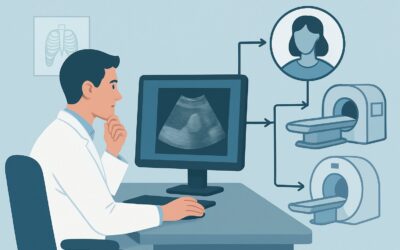
A 48-year-old woman presents for follow-up on a palpable lump in her right breast. You ordered a diagnostic mammogram with tomosynthesis, and the report is now in the EMR: a 2-cm spiculated mass at the site of the palpable concern, assessed as BI-RADS 5, highly suggestive of malignancy. The patient is scheduled to see you this afternoon, and she is understandably anxious. Your immediate task is to determine the correct next step in the diagnostic pathway. While tissue sampling is the ultimate goal, what is the appropriate imaging study to order now to characterize the lesion and prepare for biopsy?
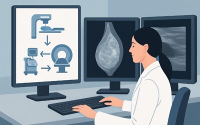
This clinical workflow article addresses this specific scenario, guided by the American College of Radiology (ACR) Appropriateness Criteria. For a woman aged 40 or older with a palpable mass and a mammogram that is suspicious or highly suggestive of malignancy (BI-RADS 4 or 5), the next recommended imaging study is a breast ultrasound, which the ACR rates as Usually Appropriate.
Who Fits This Clinical Scenario?
This guidance is specifically for an adult female, 40 years of age or older, who presents with two key findings: a palpable breast mass and a diagnostic mammogram that has already been interpreted as BI-RADS 4 (Suspicious) or BI-RADS 5 (Highly Suggestive of Malignancy). The clinical question is not about initial imaging, but about the immediate next step after a suspicious mammogram has been obtained.
This workflow is distinct from several related clinical situations:
- Initial Imaging: This article does not apply to the initial workup of a palpable mass where no imaging has yet been performed. For that scenario in a woman over 40, diagnostic mammography and ultrasound are often performed concurrently.
- Negative or Benign Mammogram: If the mammogram was negative (BI-RADS 1) or benign (BI-RADS 2) at the site of the palpable finding, the diagnostic algorithm is different, and ultrasound becomes the primary problem-solving tool.
- Probably Benign Mammogram: This guidance does not apply if the mammogram was deemed probably benign (BI-RADS 3). Those cases typically proceed to short-interval imaging follow-up rather than immediate biopsy.
- Patients Younger Than 40: The imaging workup for palpable masses in younger women (especially under 30) typically begins with ultrasound, not mammography, due to the increased density of breast tissue in that age group.
This article focuses exclusively on the patient whose workup has already crossed a critical threshold, with mammographic findings indicating a high likelihood of cancer.
What Diagnoses Are You Working Up in This Scenario?
With a BI-RADS 4 or 5 mammographic finding, the differential diagnosis is heavily weighted toward malignancy. The purpose of the subsequent ultrasound is to further characterize the lesion, assess for additional disease, and plan for tissue acquisition.
The primary consideration is invasive breast carcinoma. The most common histologic type is Invasive Ductal Carcinoma (IDC), which often forms the spiculated, irregular masses seen on mammography. A less common but important type is Invasive Lobular Carcinoma (ILC), which can be more subtle, sometimes presenting as architectural distortion rather than a discrete mass. Ultrasound is critical for identifying a targetable lesion in ILC, which can be mammographically underestimated.
Another possibility is Ductal Carcinoma In Situ (DCIS) with an associated invasive component. While pure DCIS often presents as calcifications on mammography, a palpable mass associated with it raises strong suspicion for an underlying invasive cancer. Ultrasound helps to identify the solid, invasive portion of the tumor, which is the necessary target for an accurate biopsy.
While less common, other malignancies can present this way. A malignant phyllodes tumor can appear as a large, well-circumscribed mass that may still warrant a BI-RADS 4 assessment. Rarely, primary breast lymphoma or metastases from another cancer (like melanoma or lung cancer) can manifest as a palpable, suspicious breast mass.
Finally, some benign entities can mimic malignancy on mammography, such as a radial scar (complex sclerosing lesion) or, in some cases, fat necrosis. However, given a BI-RADS 4 or 5 designation, these are considered diagnoses of exclusion that can only be confirmed by pathology after a biopsy.
Why Is Breast Ultrasound the Recommended Next Study for This Presentation?
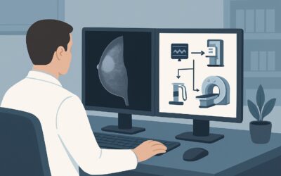
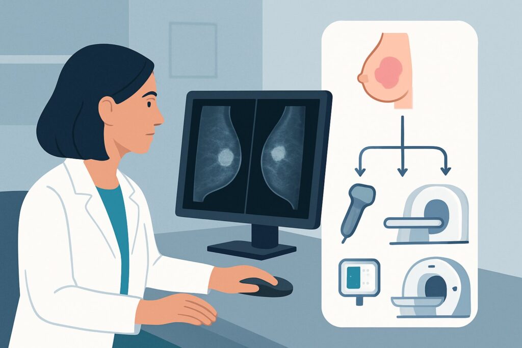
For a patient with a palpable mass and a BI-RADS 4 or 5 mammogram, breast ultrasound is rated as Usually Appropriate by the ACR because it serves several critical functions that no other imaging modality can accomplish as effectively at this stage.
The primary role of ultrasound is to provide a clear, real-time target for percutaneous biopsy. It confirms the mammographic finding corresponds to a solid mass, precisely defines its size and shape, and evaluates its margins and vascularity with Doppler. This detailed characterization is essential for planning the biopsy. Ultrasound-guided core needle biopsy is the standard of care for obtaining tissue from suspicious solid breast masses due to its high accuracy, safety, and patient tolerance.
Critically, ultrasound is also used to perform a comprehensive evaluation of the ipsilateral axilla. Detecting and sampling suspicious-looking axillary lymph nodes at the time of the initial breast biopsy is a crucial step in clinical staging. This information directly impacts subsequent surgical and systemic therapy planning.
In contrast, other imaging modalities are considered Usually Not Appropriate as the next step in this specific scenario:
- Breast MRI with and without IV contrast: While breast MRI is a highly sensitive tool for detecting cancer, its role is typically in staging after a biopsy has already confirmed malignancy. Using it before a tissue diagnosis can introduce ambiguity by identifying additional, smaller lesions that may also require biopsy, unnecessarily complicating and delaying the diagnosis of the primary, palpable lesion.
- Image-guided core biopsy: This is a procedure, not a diagnostic imaging study. The ACR’s rating clarifies that one does not simply order a “biopsy” in isolation. The preceding imaging study—ultrasound—is what determines how that biopsy will be guided. Therefore, ordering ultrasound is the correct prerequisite step.
From a safety perspective, ultrasound is ideal. It involves no ionizing radiation (0 mSv) and does not require intravenous contrast, eliminating risks of allergic reaction or contrast-induced nephropathy.
What’s Next After Breast Ultrasound? The Downstream Workflow
The results of the breast ultrasound will dictate the immediate next steps, which are almost always procedural. The workflow is designed to move efficiently toward a definitive tissue diagnosis.
If the ultrasound confirms a suspicious solid mass corresponding to the mammographic and palpable finding, the next step is an ultrasound-guided core needle biopsy. This procedure should be scheduled promptly. If the ultrasound also identifies abnormal-appearing axillary lymph nodes (e.g., cortically thickened, rounded, loss of the fatty hilum), these should also be sampled via ultrasound-guided fine-needle aspiration (FNA) or core biopsy during the same appointment.
In the rare event that a targeted ultrasound cannot identify a correlate for a clear BI-RADS 4 or 5 mammographic finding (e.g., architectural distortion or calcifications without a mass), this is considered a discordant finding. The patient should not be reassured or sent to follow-up. The next step is to proceed with a biopsy guided by the modality that best visualizes the lesion, which would be a stereotactic (mammogram-guided) biopsy.
Once the biopsy results are available and malignancy is confirmed, the patient’s care transitions to a multidisciplinary team. Further staging workup is then considered. This may include a breast MRI to evaluate the extent of disease, screen the contralateral breast, and guide surgical planning. Depending on the cancer’s stage and subtype, systemic staging with CT scans of the chest, abdomen, and pelvis, and/or a bone scan may be indicated.
Pitfalls to Avoid (and When to Get Help)
Navigating this high-stakes clinical scenario requires careful attention to detail to avoid common missteps that can delay diagnosis or lead to incomplete information.
- Pitfall 1: Incomplete Ultrasound. Do not allow a “limited” or “targeted” ultrasound of only the palpable lump. A comprehensive diagnostic breast ultrasound, including a thorough survey of all four quadrants and the entire axilla, is required to check for multicentric disease or nodal involvement.
- Pitfall 2: Dismissing a Suspicious Mammogram. If the mammogram is BI-RADS 4 or 5 but the ultrasound of the palpable area is reported as “negative” or “unremarkable,” do not stop the workup. This is a critical discordance. The mammographic finding must be addressed, typically with a stereotactic-guided biopsy.
- Pitfall 3: Delaying Tissue Sampling. Once a BI-RADS 4 or 5 lesion is identified, time is of the essence. The workflow should be streamlined to get the patient to an image-guided biopsy as quickly as possible.
If there is any discordance between the clinical exam, mammography, and ultrasound findings, it is crucial to escalate. A direct conversation with the interpreting breast radiologist is the best way to resolve ambiguity and ensure the most appropriate next step is taken.
Related ACR Topics and Tools
For a comprehensive overview of all clinical variants related to palpable breast masses, from initial workup to post-procedural follow-up, please see our parent topic guide. Additional tools from GigHz can help you apply these standards in your practice.
- For breadth across all scenarios in Palpable Breast Masses, see our parent guide: Palpable Breast Masses: ACR Appropriateness Decoded.
- To explore other clinical scenarios, use the ACR Appropriateness Criteria Lookup.
- For details on imaging techniques, visit the Imaging Protocol Library.
- To discuss cumulative radiation exposure with patients, consult the Radiation Dose Calculator.
Frequently Asked Questions
Why not go straight to an image-guided biopsy without the ultrasound?
The ultrasound is not just a preliminary step; it is the essential guidance system for the biopsy. It confirms the target, assesses its characteristics, evaluates the surrounding tissue and axillary lymph nodes for other disease, and provides the real-time visualization needed to accurately place the biopsy needle. Ordering a ‘biopsy’ without specifying the imaging guidance is incomplete; ultrasound is the study that enables the most common and effective form of biopsy for a palpable mass.
If breast MRI is so sensitive for cancer, why is it ‘Usually Not Appropriate’ at this stage?
Breast MRI is primarily a staging tool, not an initial diagnostic tool for a known suspicious mass. Its high sensitivity can be a drawback before a tissue diagnosis is established, as it may detect additional, smaller lesions (‘incidentalomas’). This can lead to multiple, sometimes unnecessary, biopsies and delay the diagnosis and treatment of the main, clinically evident cancer. Once cancer is confirmed via biopsy, MRI becomes highly valuable for defining the extent of disease and planning surgery.
What if the ultrasound shows the mass is just a complicated cyst?
While a simple cyst would be a BI-RADS 2 finding, a complicated or complex cyst can sometimes raise suspicion on a mammogram, leading to a BI-RADS 4 assessment. If ultrasound clearly characterizes the lesion as a cyst (even a complicated one) and it accounts for the mammographic finding, the radiologist may recommend aspiration or short-term follow-up instead of core biopsy. However, any solid component within a cystic structure requires biopsy.
Does this guidance change if the patient has dense breasts?
No, the guidance remains the same. In fact, ultrasound is even more critical in women with dense breasts. Mammographic sensitivity is reduced in dense tissue, making ultrasound essential for characterizing a palpable mass that may be partially obscured on the mammogram. The BI-RADS 4 or 5 assessment already indicates a suspicious finding was identified despite any density, and ultrasound is the best next step to evaluate it and the surrounding dense tissue.
The mammogram report mentions suspicious calcifications but no discrete mass. Is ultrasound still the next step?
If the suspicious finding is calcifications alone, without an associated palpable or mammographic mass, ultrasound may not be able to visualize them. In that specific case, the next step is typically a stereotactic (mammogram-guided) biopsy, which uses mammographic images to target the calcifications. However, since this scenario specifies a palpable mass, an ultrasound is still required to evaluate the palpable finding and the axilla, even if the primary mammographic abnormality is calcifications.
Reviewed by Pouyan Golshani, MD, Interventional Radiologist — May 30, 2026