What Is the Next Step for Suspected Elbow Fracture with Normal Initial X-Rays?
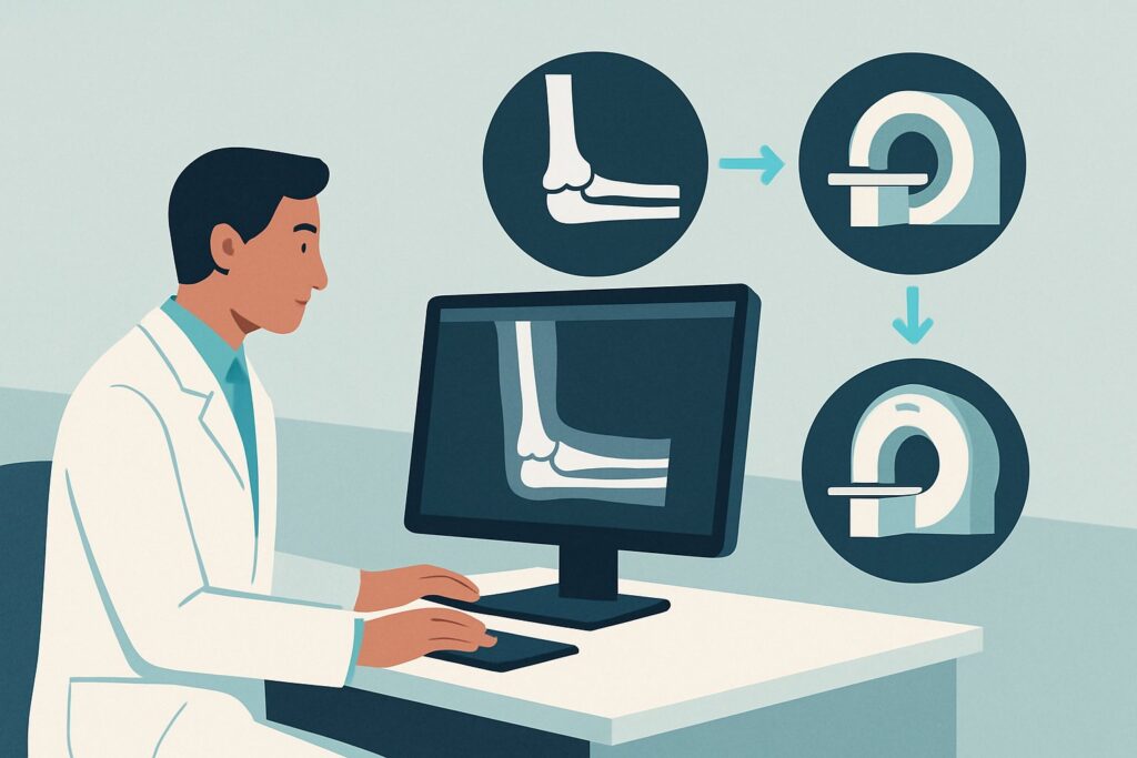
A 48-year-old office worker presents to the urgent care clinic after a fall onto their outstretched hand. They have significant pain, swelling, and point tenderness over the radial head. You order initial elbow radiographs, but the report comes back negative for acute fracture, though the radiologist notes a subtle posterior fat pad sign. Your clinical suspicion for an occult fracture remains high. What is the most appropriate next step in the imaging workup to confirm or exclude the diagnosis without ordering unnecessary or low-yield studies? This article provides a detailed workflow for this specific scenario, guided by the American College of Radiology (ACR) Appropriateness Criteria, which rate Radiography area of interest repeat in 10-14 days as Usually Appropriate.
Who Fits This Clinical Scenario?
This guidance applies to a specific patient population: an adult with acute elbow or forearm pain following trauma, where there is a strong clinical suspicion for a fracture despite normal or indeterminate initial radiographs.
Inclusion criteria for this workflow:
- Adult patient: This guidance does not apply to pediatric patients, where considerations for physeal (growth plate) injuries change the diagnostic algorithm.
- Acute traumatic injury: The pain is linked to a specific event, such as a fall or direct blow.
- High clinical suspicion for fracture: Key signs include focal bony tenderness (e.g., over the radial head or olecranon), significant swelling, ecchymosis, or an inability to fully extend the elbow.
- Negative or indeterminate initial radiographs: Standard X-rays have been performed and show no clear fracture line. An indeterminate finding might include a visible joint effusion (e.g., a positive fat pad sign) without a visible fracture.
This workflow is NOT for:
- Patients needing initial imaging: If no imaging has been performed yet, the correct scenario is “Adult. Acute elbow or forearm pain. Initial imaging.”
- Suspected primary soft tissue injury: If the primary clinical concern is a tendon rupture (e.g., distal biceps), ligament sprain, or muscle tear, the imaging pathway is different. This routes to the ACR variant for suspected tendon or ligament injury.
- Obvious fractures on initial X-rays: If a fracture is clearly visible on the first set of images, the diagnostic question is answered, and the focus shifts to orthopedic management.
What Diagnoses Are You Working Up in This Scenario?
When initial radiographs are negative in the setting of trauma and high clinical suspicion, you are primarily searching for an occult fracture—one that is not yet visible on standard X-rays. The differential diagnosis is focused on common but subtle bony injuries.
The most frequent culprit in this scenario is an occult radial head fracture. This is a classic injury following a fall on an outstretched hand (FOOSH). These fractures are often non-displaced or only minimally displaced, making the fracture line nearly impossible to see on initial films. The presence of an anterior or posterior fat pad sign on a lateral radiograph is a sensitive indirect indicator of a joint effusion, which in the setting of trauma is presumed to be a hemarthrosis from an occult fracture until proven otherwise.
A less common but important consideration is an occult olecranon or coronoid process fracture. Olecranon fractures typically result from a direct blow to the posterior elbow, while coronoid process fractures are often associated with more complex elbow instability or dislocation events. Both can be difficult to visualize if non-displaced.
Finally, a significant bone bruise (trabecular microfracture) can present with the same clinical signs as a true cortical fracture. While not a true fracture in the traditional sense, a bone bruise involves bleeding and edema within the bone marrow and can be a source of significant pain and disability. This diagnosis is typically made with advanced imaging like MRI but is part of the differential for severe post-traumatic pain with negative radiographs.
Why Is Repeating Radiographs in 10-14 Days a Recommended Study?
For a patient with a suspected occult elbow fracture, the ACR panel rates Radiography area of interest repeat in 10-14 days as Usually Appropriate. This strategy leverages basic bone biology to achieve a diagnosis in a cost-effective and low-radiation manner. After a fracture, the initial inflammatory response and subsequent bone resorption at the fracture margins cause the fracture line to widen slightly. This process makes a previously invisible fracture line become visible on radiographs taken 10 to 14 days after the initial injury.
While waiting is the traditional and often best approach, CT area of interest without IV contrast is also rated Usually Appropriate. CT provides exquisite bony detail and can identify an occult fracture immediately. It is the preferred next step for patients who require a definitive and rapid diagnosis, such as high-performance athletes, laborers who need to know their return-to-work timeline, or in cases where a complex intra-articular fracture pattern is suspected. The trade-off is higher cost and a greater radiation dose (RRL: Varies) compared to repeat radiographs.
Alternative studies are rated lower for this specific indication:
- MRI area of interest without IV contrast is rated Usually not appropriate. While MRI is extremely sensitive for detecting bone marrow edema and can confirm a bone bruise or occult fracture with near-perfect accuracy, it is often diagnostic overkill for this simple question. It is more expensive, takes longer to acquire, and is less readily available than CT or radiography. Its primary role is reserved for cases where a significant ligamentous or soft tissue injury is the main concern, not an isolated occult fracture.
- US area of interest (ultrasound) is also rated Usually not appropriate. Ultrasound is excellent for evaluating soft tissues, tendons, and fluid collections, but it has limited utility for diagnosing occult fractures of the elbow in adults. Visualizing the complete bone cortex, especially the radial head, is difficult and highly operator-dependent.
The decision between watchful waiting with repeat radiographs and proceeding directly to CT depends on clinical judgment, patient factors, and the urgency of establishing a definitive diagnosis.
What’s Next After Repeat Radiographs? Downstream Workflow
The results of the follow-up study will guide your next steps in management. This decision tree outlines the most common pathways.
- If the repeat radiograph is POSITIVE for a fracture: The diagnosis is confirmed. For most non-displaced or minimally displaced radial head fractures (the most common finding), management is conservative with a brief period of immobilization (sling) followed by early range of motion exercises. Refer the patient to an orthopedic specialist for definitive management recommendations, as even simple fractures can lead to stiffness if not managed properly.
- If the repeat radiograph is NEGATIVE but the patient remains symptomatic: If the patient continues to have significant pain, mechanical symptoms (like locking or clicking), or an inability to regain motion, the initial diagnosis of a simple sprain may be incorrect. At this point, the likelihood of a significant soft tissue injury (e.g., ligament tear) or a true occult fracture still missed by two sets of radiographs increases. This is the point where escalating to advanced imaging is warranted. The next logical step would often be an MRI without contrast to evaluate the bone marrow, cartilage, and soft tissues in detail.
- If the repeat radiograph is NEGATIVE and the patient’s symptoms are improving: This clinical course is most consistent with a minor sprain, bone bruise, or a clinically insignificant fracture that has healed without consequence. Continue conservative management with a focus on physical therapy and a gradual return to activity. No further imaging is typically required.
Pitfalls to Avoid (and When to Get Help)
Navigating this clinical scenario requires careful attention to subtle signs and patient progression. Here are a few common pitfalls to avoid:
- Dismissing the fat pad sign: A posterior fat pad sign or a large, bulging anterior fat pad sign on a lateral elbow X-ray is never normal in an adult. In the context of trauma, it should be treated as an intra-articular fracture until proven otherwise.
- Prolonged immobilization: For suspected non-displaced radial head fractures, prolonged casting or splinting can lead to significant and sometimes permanent stiffness. The standard of care is often early mobilization.
- Ignoring mechanical symptoms: If a patient reports locking, catching, or instability, even with negative radiographs, there may be an intra-articular loose body or a complex ligamentous injury. This warrants a lower threshold for advanced imaging (CT or MRI) and orthopedic consultation.
If a patient develops neurological symptoms (numbness, tingling), vascular compromise (cool, pale hand), or shows signs of a compartment syndrome (pain out of proportion, tense forearm), this constitutes a clinical emergency requiring immediate escalation to an orthopedic surgeon or the emergency department.
Related ACR Topics and Tools
This article covers one specific variant within the broader topic of elbow and forearm pain. For a comprehensive overview and to explore related clinical scenarios, please consult the resources below.
- For breadth across all scenarios in Acute Elbow and Forearm Pain, see our parent guide: Acute Elbow and Forearm Pain: ACR Appropriateness Decoded.
- ACR Appropriateness Criteria Lookup — for adjacent scenarios
- Imaging Protocol Library — for technique on the recommended study
- Radiation Dose Calculator — for cumulative dose conversations
Frequently Asked Questions
Why wait 10-14 days for a repeat X-ray instead of getting a CT scan right away?
Waiting 10-14 days allows for biological changes at the fracture site (bone resorption) to make a subtle fracture line more visible on a simple radiograph. This is a highly effective, low-cost, and low-radiation strategy. A CT scan is also appropriate and provides an immediate answer, but it involves more radiation and expense. The choice depends on the clinical urgency and patient-specific factors.
What is the significance of a ‘fat pad sign’ on the initial elbow X-ray?
In adults, a visible posterior fat pad or a prominent ‘sail-like’ anterior fat pad on a lateral elbow radiograph indicates a joint effusion. In the setting of acute trauma, this effusion is presumed to be blood (a hemarthrosis) from an intra-articular fracture, most commonly of the radial head. It is a strong indirect sign of an occult fracture.
If the repeat radiograph is negative but the patient still has significant pain, what is the next step?
If high clinical suspicion for a significant injury persists despite two sets of negative radiographs, advanced imaging is warranted. An MRI without contrast is typically the best next step, as it can evaluate for bone bruises (marrow edema), cartilage injury, and ligament or tendon tears that would not be visible on X-ray or CT.
Is an MRI better than a CT for finding an occult elbow fracture?
Both are highly sensitive for occult fractures. However, CT is generally preferred if the only question is confirming a bony fracture, as it provides superior cortical bone detail quickly and cost-effectively. MRI is better if you also suspect a significant soft tissue injury (ligament, tendon, cartilage) or want to confirm a bone bruise, but it is rated ‘Usually not appropriate’ by the ACR for the sole purpose of finding a simple occult fracture.
Should I immobilize the elbow while waiting for the repeat radiograph?
Yes, if an occult fracture is suspected, it is appropriate to treat the injury as a fracture. This typically involves placing the arm in a sling and/or a posterior splint for comfort and protection for the 10-14 day waiting period. Advise the patient to avoid weight-bearing and forceful activities with the injured arm.
Reviewed by Pouyan Golshani, MD, Interventional Radiologist — May 30, 2026