What Is the Right First Imaging Study for an Adult with Chronic Hand or Wrist Pain?
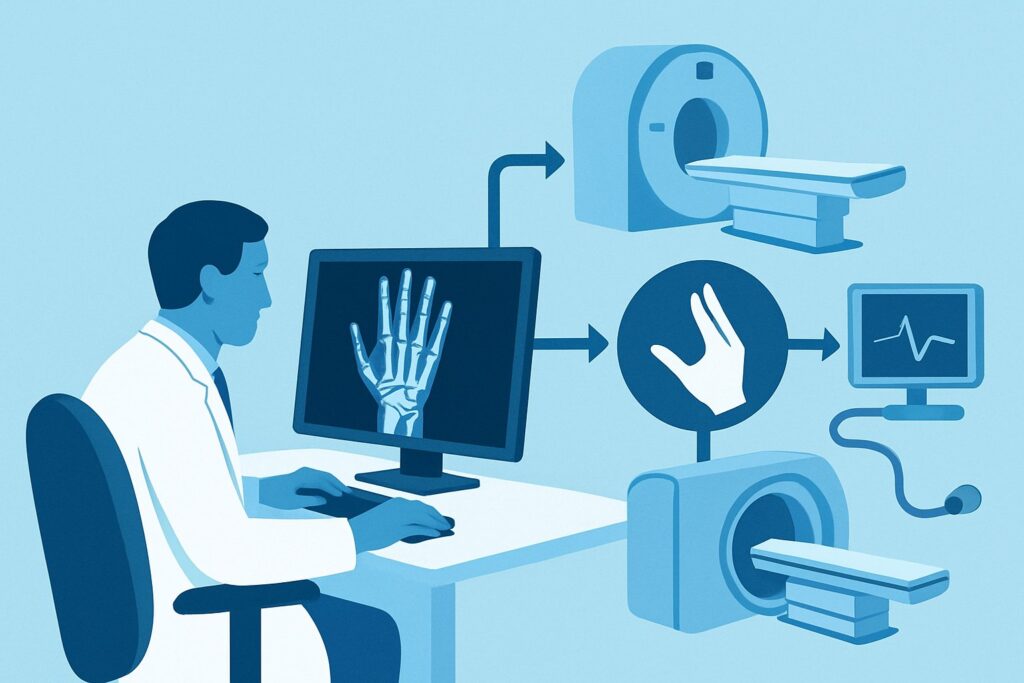
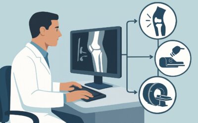
A 48-year-old administrative assistant presents to your clinic with four months of progressive, aching pain in her dominant wrist. There was no specific injury, but the pain is now constant, aggravated by typing and lifting. On exam, there is mild swelling and tenderness over the radiocarpal joint. You are at the electronic health record, ready to order the initial imaging study to begin the diagnostic workup. For this common clinical presentation—an adult with chronic, non-traumatic hand or wrist pain—the American College of Radiology (ACR) provides clear guidance to ensure an efficient and effective evaluation. The foundational first step, designated as Usually appropriate, is radiography of the area of interest. This article details the clinical workflow for this specific scenario, explaining the rationale for this choice and outlining the next steps based on the findings.
Who Fits This Clinical Scenario for Chronic Hand or Wrist Pain?
This guidance applies to a specific and common patient population: an adult presenting with hand or wrist pain that has persisted for more than six weeks without a history of significant, acute trauma. The key elements are the chronicity of the symptoms and the absence of a clear inciting event. The pain is the primary complaint, and the goal of imaging is to establish a diagnosis for these undifferentiated symptoms.
It is equally important to recognize who does not fit this scenario. This workflow is not intended for patients where:
- Previous radiographs are already available and normal. If you have recent, normal x-rays, the clinical question has advanced. The workup would then follow the ACR variant for “Chronic wrist pain. Radiographs normal or remarkable for nonspecific arthritis. Next imaging study.”
- Symptoms are highly specific for carpal tunnel syndrome. If the patient’s primary complaints are numbness and tingling in the median nerve distribution, especially at night, the evaluation shifts to the specific ACR criteria for suspected carpal tunnel syndrome, which may involve nerve conduction studies or specific imaging protocols.
- There is a known prior fracture. A patient with a history of a scaphoid fracture and persistent pain requires a different workup focused on evaluating for nonunion, malunion, or osteonecrosis.
- A tendon injury is strongly suspected. If the history and exam point specifically to a tendon issue, such as triggering, locking, or a palpable defect, the imaging choice may be different, often favoring ultrasound.
Correctly identifying your patient’s scenario is the first step to an efficient diagnostic pathway.
What Diagnoses Are You Working Up With Initial Imaging?
When ordering the first imaging study for chronic hand or wrist pain, the goal is to evaluate for the most common underlying causes, most of which involve the osseous structures. The differential diagnosis is broad, but radiography is an excellent tool for assessing several key possibilities.
The most frequent cause of chronic wrist pain in adults is osteoarthritis (OA), or degenerative joint disease. Radiographs are highly effective at identifying the classic signs of OA, including joint space narrowing, the formation of osteophytes (bone spurs), and increased density of the subchondral bone.
Next are the inflammatory arthritides, such as rheumatoid arthritis (RA) or psoriatic arthritis. While early findings can be subtle, radiographs can reveal characteristic signs like soft tissue swelling, periarticular osteopenia, and, in more advanced cases, marginal erosions and symmetric joint space loss. These findings can be crucial in prompting a rheumatologic consultation.
Less common but important to exclude is avascular necrosis (AVN), a condition where blood supply to a bone is disrupted, leading to bone death. In the wrist, this most often affects the lunate (Kienböck disease) or the scaphoid (Preiser disease). Radiographs can show increased bone density (sclerosis), cystic changes, or eventual collapse of the affected carpal bone, though they are insensitive in the earliest stages.
Finally, radiographs can identify evidence of crystal deposition diseases like gout or calcium pyrophosphate deposition (CPPD) disease. Gout may present with characteristic “punched-out” erosions with overhanging edges, while CPPD can be identified by the presence of chondrocalcinosis—calcification within the cartilage, particularly of the triangular fibrocartilage complex (TFCC).
Why Is Radiography the Recommended First Study for Chronic Hand and Wrist Pain?
The ACR Appropriateness Criteria rate Radiography area of interest as Usually appropriate for the initial evaluation of an adult with chronic hand or wrist pain. This recommendation is based on the modality’s high diagnostic yield for common pathologies, wide availability, low cost, and favorable safety profile. It serves as the essential first-line test that effectively screens for the most prevalent osseous and articular abnormalities.
Radiography provides an excellent overview of bone and joint alignment, joint space integrity, and bone texture. It is the most direct and efficient way to diagnose or exclude osteoarthritis, many inflammatory conditions, and advanced avascular necrosis. Even when the radiographs are normal, they provide a critical baseline and help narrow the differential, guiding the decision for any subsequent, more advanced imaging.
In contrast, other more advanced modalities are rated lower for this initial step:
- MRI without IV contrast is rated Usually not appropriate. While MRI offers superior detail of soft tissues and early bone marrow changes, it is significantly more expensive and less accessible. Ordering it as the first test bypasses the valuable information provided by a simple radiograph and represents an inefficient use of resources for an undifferentiated presentation. It is best reserved as a problem-solving tool when radiographs are negative but clinical suspicion for a specific condition remains high.
- Ultrasound of the area of interest is rated May be appropriate. Ultrasound excels at evaluating soft tissues like tendons, ligaments, and synovium and can detect fluid collections and synovitis with high sensitivity. However, it is highly operator-dependent and provides a limited assessment of the underlying bone structure. While it can be a valuable adjunct or a primary tool for specific soft-tissue concerns, radiography remains the superior initial screening test for undifferentiated chronic pain.
From a safety perspective, radiography of an extremity involves a low level of ionizing radiation (ACR Relative Radiation Level: Varies). This is a stark contrast to a modality like a nuclear medicine bone scan, which is rated Usually not appropriate for this scenario and carries a significantly higher radiation dose (☢☢☢ 1-10 mSv).
What’s the Next Step After Initial Radiographs?
The results of the initial radiographs create a critical branch point in the clinical workflow. The next step is determined entirely by the findings in the context of the patient’s symptoms.
If the radiographs are positive and explain the symptoms:
For many patients, the radiographs will reveal clear evidence of osteoarthritis or another condition that correlates well with their clinical presentation. In these cases, the diagnostic workup may be complete. The focus shifts to management, which can include activity modification, splinting, anti-inflammatory medications, or referral to physical or occupational therapy. Further imaging is typically not required unless the patient fails to respond to conservative treatment or develops new symptoms.
If the radiographs are negative or show nonspecific findings:
A normal radiograph is a common and important result. It effectively rules out significant degenerative disease, advanced AVN, and many fractures. However, it does not exclude soft-tissue pathology, early inflammatory arthritis, or the early stages of AVN. At this point, the workup transitions to a new clinical scenario: “Chronic hand or wrist pain with normal radiographs.” The next step, guided by the specific clinical suspicion, is often advanced imaging.
- If an occult fracture, early AVN, or internal derangement is suspected, an MRI is typically the next best test.
- If tenosynovitis or a tendon injury is the leading concern, a diagnostic ultrasound may be more appropriate.
If the radiographs suggest inflammatory arthritis or a complex issue:
Findings such as erosions, characteristic joint space loss, or signs of AVN warrant a specialist referral. A rheumatologist should be consulted for suspected inflammatory arthritis, while a hand surgeon is the appropriate consultant for conditions like Kienböck disease or complex post-traumatic changes. These specialists will guide any further imaging and management.
Common Pitfalls to Avoid in This Initial Workup
Navigating the initial workup for chronic hand and wrist pain requires careful clinical correlation to avoid common missteps.
- Stopping the workup after a “negative” x-ray. A normal radiograph is not the end of the investigation if the patient remains symptomatic. It rules out major bone and joint disease but should prompt consideration of soft-tissue causes.
- Ordering inadequate radiographic views. Standard posteroanterior (PA), lateral, and oblique views are essential. If there is high suspicion for a specific problem, such as scaphoid pathology, be sure to order dedicated views (e.g., a scaphoid view) to increase diagnostic sensitivity.
- Jumping to advanced imaging. Ordering an MRI as the first test is rarely the right answer. It is less cost-effective and bypasses the crucial foundational information that a simple radiograph provides, potentially leading to the discovery of incidental findings that complicate the clinical picture.
A critical exception to this workflow is the presence of red flag symptoms. If you suspect an infection (fever, erythema, warmth), a tumor (palpable mass, constitutional symptoms), or acute neurovascular compromise, you should escalate immediately to more advanced imaging, typically an MRI, and obtain an urgent specialist consultation.
Related ACR Topics and Tools
The ACR Appropriateness Criteria are a comprehensive resource for evidence-based imaging decisions. For a broader overview of all clinical variants related to this topic, or to explore the tools used to apply this guidance in practice, please see the resources below.
For breadth across all scenarios in Chronic Hand and Wrist Pain, see our parent guide: Chronic Hand and Wrist Pain: ACR Appropriateness Decoded.
- ACR Appropriateness Criteria Lookup — for adjacent scenarios
- Imaging Protocol Library — for technique on the recommended study
- Radiation Dose Calculator — for cumulative dose conversations
Frequently Asked Questions
Why not just order an MRI first for chronic wrist pain to see everything at once?
While MRI provides excellent detail, the ACR rates it ‘Usually not appropriate’ as a first step. Radiographs are highly effective for diagnosing the most common causes of chronic wrist pain, like osteoarthritis, are less expensive, and more widely available. A radiograph provides an essential baseline that helps determine if an MRI is even needed, making the overall diagnostic process more efficient.
What specific radiographic views should I order for undifferentiated wrist pain?
A standard three-view series, including posteroanterior (PA), lateral, and oblique views, is the recommended starting point. If your clinical exam reveals focal tenderness over a specific area, such as the anatomic snuffbox, you should also order dedicated views (e.g., a scaphoid view) to increase sensitivity for subtle fractures.
Is ultrasound a reasonable alternative to radiographs as a first imaging test?
The ACR rates ultrasound as ‘May be appropriate.’ It is an excellent tool for evaluating soft tissues, such as tendons and synovium, and can be useful if your clinical suspicion points strongly to a condition like tenosynovitis. However, for general, undifferentiated chronic pain, radiography is the superior initial test because it provides a more comprehensive view of the bones and joints, which are the source of the most common pathologies.
If a patient had normal wrist x-rays several years ago, do I need to repeat them?
Yes. For a new or persistent presentation of chronic pain, current radiographs are essential. Chronic conditions like osteoarthritis are progressive, and new findings may have developed since the prior study. A new set of radiographs establishes an up-to-date baseline for comparison and is a mandatory step before considering more advanced imaging.
Does this guidance apply if the patient recalls a minor injury months ago?
Yes. If the presentation has evolved from an acute injury to a state of chronic, persistent pain, this workflow for initial imaging is the correct approach. The goal is to evaluate for underlying causes of chronic symptoms, such as post-traumatic arthritis or nonunion of a previously unrecognized fracture, for which radiography is the appropriate first step.
Reviewed by Pouyan Golshani, MD, Interventional Radiologist — May 30, 2026