What’s the Best Initial Imaging for Suspected Occupational Interstitial Lung Disease?
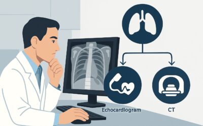
A 55-year-old man, a retired stonemason with over 30 years of exposure to silica dust, presents to your clinic with a six-month history of progressive shortness of breath and a persistent dry cough. His pulmonary function tests show a restrictive pattern. You suspect an occupational interstitial lung disease (ILD), but the differential is broad. Your immediate question is what imaging to order first to confirm your suspicion and guide the next steps in management. This article provides a detailed clinical workflow for this specific scenario, grounded in the American College of Radiology (ACR) Appropriateness Criteria. For the initial imaging of a patient with occupational exposure and suspected ILD, a chest radiograph is rated Usually Appropriate.
Who Fits This Clinical Scenario?
This guidance is for patients presenting with a new or uninvestigated clinical suspicion of interstitial lung disease coupled with a significant history of occupational exposure. The key inclusion criteria are:
- A documented or strongly suspected history of meaningful exposure to fibrogenic dusts or agents (e.g., silica, asbestos, coal dust, beryllium, organic antigens).
- Clinical signs and symptoms suggestive of ILD, such as progressive dyspnea on exertion, persistent non-productive cough, or inspiratory crackles on auscultation.
- This is the initial imaging workup for this specific presentation; the patient has not had prior thoracic imaging for this problem.
This workflow is distinct from related but different clinical situations. This article does not apply if:
- The patient is asymptomatic: The evaluation of an asymptomatic but exposed worker falls under the screening and surveillance guidelines, which have a different risk-benefit calculus.
- The primary suspicion is airway disease: If symptoms like wheezing and productive cough dominate, suggesting occupational asthma or chronic bronchitis, the imaging approach is different.
- A chest radiograph has already been performed: If a radiograph has already identified findings concerning for ILD, the question is no longer about initial imaging but about the appropriate next study, a separate ACR scenario.
What Diagnoses Are You Working Up in This Scenario?
When ordering initial imaging for suspected occupational ILD, you are primarily investigating a group of diseases known as the pneumoconioses, while also considering important mimics. The imaging findings will help narrow this differential.
Pneumoconioses: This is the core diagnostic category. These are non-neoplastic lung reactions to inhaled mineral or organic dusts. The most common are silicosis (from silica dust, seen in mining, quarrying, and sandblasting), asbestosis (from asbestos fibers, seen in construction and shipbuilding), and Coal Worker’s Pneumoconiosis (CWP). Imaging helps identify characteristic patterns, such as upper-lobe predominant nodules in silicosis or basilar reticulation and pleural plaques in asbestosis.
Hypersensitivity Pneumonitis (HP): While also occupation-related, HP is an immunologic reaction to inhaled organic antigens, such as mold from hay (“Farmer’s Lung”) or avian proteins (“Bird Fancier’s Lung”). It can present with ground-glass opacities, centrilobular nodules, and, in chronic forms, fibrosis that can mimic other ILDs.
Idiopathic Pulmonary Fibrosis (IPF): A crucial and more common diagnosis to differentiate from occupational ILDs, particularly asbestosis. IPF is a progressive fibrotic lung disease of unknown cause. While the occupational history is the key clinical differentiator, the imaging pattern of usual interstitial pneumonia (UIP) with basal and subpleural predominant honeycombing can sometimes overlap with asbestosis, making careful radiologic interpretation essential.
Sarcoidosis: This is a systemic granulomatous disease of unknown etiology that can present with respiratory symptoms and radiographic findings—such as bilateral hilar lymphadenopathy and reticulonodular infiltrates—that can mimic silicosis or berylliosis. Distinguishing these is critical as management and prognosis differ significantly.
Why Is a Chest Radiograph the Recommended Initial Study?
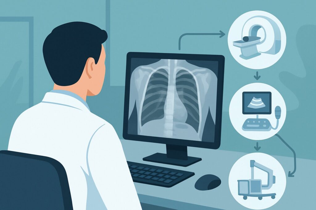
The ACR rates both chest radiography and non-contrast chest CT as Usually Appropriate for the initial evaluation of suspected occupational ILD. However, the standard clinical workflow begins with the chest radiograph due to its unique combination of diagnostic utility, accessibility, and safety for this specific question.
A chest radiograph is an excellent first-line tool. It is widely available, inexpensive, and quick to perform. Critically, it provides a very low radiation dose (ACR Relative Radiation Level ☢, <0.1 mSv), which is an important consideration in any initial diagnostic pathway. Despite the superior detail of CT, a standard two-view chest radiograph is often sufficient to detect the classic macroscopic findings of many pneumoconioses. It can readily identify diffuse reticular or nodular patterns, pleural plaques or thickening characteristic of asbestos exposure, and the “egg-shell” calcification of hilar lymph nodes sometimes seen in silicosis.
While non-contrast chest CT is also Usually Appropriate and is the gold standard for characterizing ILD, it is typically reserved as the next step after an abnormal or inconclusive radiograph. Starting with CT exposes the patient to a significantly higher radiation dose (ACR Relative Radiation Level ☢☢☢, 1-10 mSv) without first establishing a clear need for its superior resolution.
Other imaging modalities are rated lower for this initial workup for clear reasons:
- CT Chest with IV Contrast is rated Usually Not Appropriate. Intravenous contrast is used to evaluate vascular structures, mediastinal masses, or active inflammation. It offers no additional value for assessing the fine structural details of the lung parenchyma in suspected ILD and adds the risks of contrast reaction and nephrotoxicity.
- MRI of the Chest is also Usually Not Appropriate. MRI has poor spatial resolution for the delicate lung interstitium compared to CT and is susceptible to motion artifacts from breathing and cardiac motion, making it unsuitable for diagnosing or characterizing ILD.
What’s Next After a Chest Radiograph? Downstream Workflow
The results of the initial chest radiograph will dictate the subsequent diagnostic pathway. The goal is to move efficiently toward a confident diagnosis, which often requires more advanced imaging.
If the radiograph is positive or suspicious for ILD: Any finding suggestive of interstitial disease—such as reticulation, nodules, ground-glass opacities, or pleural abnormalities—warrants further characterization. The definitive next step is a High-Resolution Computed Tomography (HRCT) scan of the chest without IV contrast. This is the subject of a distinct ACR variant: “Occupational exposure, suspected interstitial lung disease based on radiography. Next imaging study.” HRCT provides the detailed anatomical information needed to classify the pattern of fibrosis (e.g., UIP, NSIP), assess its distribution, and identify subtle findings missed on the radiograph, which is essential for narrowing the differential diagnosis.
If the radiograph is negative: A normal chest radiograph does not definitively exclude interstitial lung disease, especially in its early stages. If your clinical suspicion remains high based on the patient’s symptoms, exposure history, and restrictive PFTs, proceeding directly to a non-contrast HRCT of the chest is the appropriate next step. Early or subtle ILD can be radiographically occult but readily apparent on HRCT.
If the radiograph is indeterminate: Findings like “increased interstitial markings” or “bibasilar atelectasis” are nonspecific. In these cases, clinical correlation is key. If the patient’s symptoms and risk profile are compelling, these equivocal findings should lower the threshold to proceed to HRCT for a more definitive evaluation.
Pitfalls to Avoid (and When to Get Help)
Navigating the initial workup for occupational ILD requires careful attention to detail to avoid common missteps.
- Over-relying on a negative radiograph: Do not dismiss a strong clinical suspicion for ILD based solely on a normal chest X-ray. Early disease is often missed.
- Ordering the wrong CT: When you do proceed to CT, ensure you order a non-contrast, high-resolution (HRCT) protocol. Ordering a standard CT or a CT with contrast will not provide the thin-slice images necessary to properly evaluate the lung interstitium.
- Ignoring the exposure history: The radiologist’s interpretation is significantly enhanced by a detailed clinical history. Always specify the suspected exposure (e.g., “rule out asbestosis, history of shipyard work”) on the imaging requisition.
- Anchoring on a single diagnosis: The radiographic patterns of different ILDs can overlap. Maintain a broad differential and be prepared to pursue further workup, such as serology, bronchoscopy, or surgical lung biopsy, if imaging is not definitive.
If the imaging findings are complex or do not fit a classic pattern, consultation with a pulmonologist and/or a thoracic radiologist with expertise in diffuse lung disease is the critical next step.
Related ACR Topics and Tools
For a comprehensive overview of imaging across all related clinical presentations, please consult our parent topic guide. For specific questions about other scenarios or imaging techniques, the following resources provide direct access to authoritative guidance and data.
- For breadth across all scenarios in Occupational Lung Diseases, see our parent guide: Occupational Lung Diseases: ACR Appropriateness Decoded.
- For adjacent scenarios not covered here, use the ACR Appropriateness Criteria Lookup.
- For technical details on imaging studies, explore the Imaging Protocol Library.
- For discussions about radiation exposure with patients, see the Radiation Dose Calculator.
Frequently Asked Questions
Why is a chest radiograph recommended over a non-contrast CT when both are rated ‘Usually Appropriate’?
While both are considered appropriate, the standard clinical workflow prioritizes the chest radiograph as the initial step due to its significantly lower radiation dose, lower cost, and high availability. It is often sufficient to detect clear evidence of ILD. The non-contrast CT, while more sensitive, is typically reserved for cases where the radiograph is positive, negative but clinical suspicion remains high, or indeterminate.
What is the difference between a standard chest CT and a High-Resolution CT (HRCT)?
An HRCT uses very thin-slice images (typically 1-1.5 mm) and a specific reconstruction algorithm to provide exquisite detail of the lung parenchyma and interstitium. A standard chest CT uses thicker slices (3-5 mm), which can obscure the fine patterns of interstitial lung disease. For an ILD workup, an HRCT protocol is essential.
If I suspect asbestosis, should I order a CT first to look for pleural plaques?
Not necessarily. While CT is more sensitive for detecting pleural plaques, a standard chest radiograph is often the appropriate first test. It can frequently identify calcified and non-calcified pleural plaques, as well as parenchymal fibrosis. If the radiograph is suggestive or if clinical suspicion is high despite a negative radiograph, an HRCT is the correct next step.
Does a normal chest radiograph rule out occupational lung disease?
No. A normal chest radiograph does not exclude early or mild interstitial lung disease. Conditions like hypersensitivity pneumonitis or early asbestosis can be present with a normal radiograph. If symptoms, exposure history, and pulmonary function tests are highly suggestive of ILD, a normal radiograph should prompt further evaluation with an HRCT.
When would a CT with IV contrast be appropriate in a patient with known occupational lung disease?
A CT with IV contrast would be appropriate if you are investigating a complication or a different, superimposed condition. For example, if you suspect a pulmonary embolism, a thoracic malignancy (to assess mediastinal involvement), or an infectious complication like an abscess, IV contrast would be necessary. However, for the initial diagnosis and characterization of the ILD itself, contrast is not indicated.
Reviewed by Pouyan Golshani, MD, Interventional Radiologist — May 29, 2026