Which Imaging Is Best for Pulmonary Metastasis Staging in Musculoskeletal Tumors?
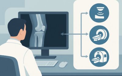
It’s late in the afternoon, and you’re coordinating care for a 17-year-old male with a new biopsy-proven osteosarcoma of the distal femur. The orthopedic oncology team is planning neoadjuvant chemotherapy, but first, complete staging is required. The most common site of distant spread for high-grade sarcomas is the lungs, and your immediate task is to order the correct imaging study to evaluate for pulmonary metastases. This decision will fundamentally alter the patient’s stage, prognosis, and treatment plan. For this specific clinical question—initial staging for lung metastases from a primary musculoskeletal tumor—the American College of Radiology (ACR) rates CT chest without IV contrast as ‘Usually Appropriate’. This article details the clinical workflow and rationale behind that recommendation.
Who Fits This Clinical Scenario?
This guidance applies to a specific and critical point in patient care: the initial staging of a newly diagnosed, biopsy-proven malignant or aggressive primary musculoskeletal tumor. The patient has not yet undergone definitive treatment, and the clinical question is focused exclusively on identifying or ruling out pulmonary metastases.
Inclusion criteria for this workflow:
- Patients with a new diagnosis of a primary bone or soft tissue sarcoma (e.g., osteosarcoma, Ewing sarcoma, rhabdomyosarcoma, undifferentiated pleomorphic sarcoma).
- The imaging is part of the initial, pre-treatment staging workup.
- The explicit goal is to evaluate the lung parenchyma for metastatic nodules.
Exclusion criteria (patients who require a different workflow):
- Evaluation for Extrapulmonary Metastasis: If you are also looking for bone, lymph node, or other distant metastases, a different imaging strategy, such as whole-body PET/CT, may be more suitable. This workflow is only for the pulmonary component of staging.
- Post-Treatment Surveillance: Patients who have completed initial therapy and are now undergoing routine follow-up to monitor for recurrence fall under a separate surveillance scenario with different imaging considerations.
- Evaluation for Local Recurrence: Assessing the primary tumor site for recurrence after surgery or radiation is a distinct clinical question, typically addressed with MRI or other modalities focused on the primary site.
What Diagnoses Are You Working Up in This Scenario?
While the primary tumor is already known, the chest imaging study is designed to differentiate between several key possibilities within the lungs. The findings will determine if the patient has localized (M0) or metastatic (M1) disease.
Pulmonary Metastases
This is the primary diagnostic target. For most high-grade sarcomas, pulmonary metastases classically appear as multiple, well-circumscribed, spherical nodules of varying sizes. They often have a peripheral and lower-lobe predominance due to hematogenous spread. Detecting these nodules, even when small, is critical for accurate staging and is the main purpose of the imaging study.
Benign Pulmonary Nodules
A significant confounder in chest imaging is the high prevalence of benign nodules from other causes, such as healed granulomas (from prior fungal or mycobacterial infection) or intrapulmonary lymph nodes. A high-quality imaging study must provide enough detail for the radiologist to confidently distinguish these common, benign findings from true metastases based on features like size, morphology, location, and calcification.
Synchronous Primary Lung Cancer
While uncommon, especially in the younger demographic typical for many primary bone sarcomas, the possibility of a second, unrelated primary lung cancer exists in older patients or those with a significant smoking history. This is a less frequent consideration but remains on the differential, as its presence would represent a separate oncologic process requiring a distinct management plan.
Why Is CT Chest Without IV Contrast the Recommended Study?
The ACR designates CT chest without IV contrast as ‘Usually Appropriate’ for this scenario because it provides the optimal balance of diagnostic accuracy, safety, and efficiency for detecting pulmonary nodules.
The rationale is grounded in physics and pathophysiology. The lung parenchyma provides excellent intrinsic contrast; dense soft-tissue nodules stand out clearly against the surrounding air-filled lung. This high natural contrast makes intravenous contrast material unnecessary for simple nodule detection. In fact, IV contrast can sometimes obscure small nodules located adjacent to pulmonary vessels, potentially reducing sensitivity.
A non-contrast CT offers exceptional spatial resolution, allowing for the detection of nodules just a few millimeters in size—far smaller than what can be reliably seen on a chest radiograph. This sensitivity is paramount, as under-staging a patient by missing small-volume metastatic disease can lead to undertreatment.
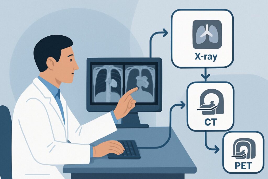
Why are other studies rated lower?
- Radiography chest is rated ‘Usually not appropriate’ due to its poor sensitivity. It can easily miss nodules smaller than 1 cm, which is unacceptable when accurate staging is the goal. A negative chest radiograph provides false reassurance and is insufficient for this clinical indication.
- CT chest with IV contrast is also ‘Usually not appropriate’. As mentioned, the contrast provides no added benefit for detecting parenchymal nodules and introduces unnecessary risks (e.g., contrast reaction, nephrotoxicity) and cost.
- FDG-PET/CT whole body is rated ‘May be appropriate’. While it is an excellent tool for evaluating for metabolically active extrapulmonary disease, it is not the primary test for pulmonary staging. Its spatial resolution is lower than a dedicated diagnostic chest CT, meaning it can miss small or sub-centimeter nodules that are not avid enough to be detected. It also carries a substantially higher radiation dose (adult RRL ☢☢☢☢, 10-30 mSv) compared to a non-contrast chest CT (adult RRL ☢☢☢, 1-10 mSv).
The radiation dose for a pediatric non-contrast chest CT (pediatric RRL ☢☢☢☢, 3-10 mSv) is a key consideration, but the diagnostic yield is considered essential for guiding therapy in a life-threatening malignancy. Once you’ve decided on the top procedure, our protocol guide covers the technique, contrast, and reading principles: CT Chest Without Contrast.
What’s Next After CT Chest Without IV Contrast? Downstream Workflow
The results of the staging chest CT will direct the subsequent steps in the patient’s management. The workflow branches based on whether the findings are clearly positive, negative, or indeterminate.
If the study is positive for definite metastases:
The presence of one or more nodules with a typical appearance for metastases confirms Stage IV disease. The patient is upstaged to M1. This finding is presented at a multidisciplinary tumor board, and the treatment plan is adjusted accordingly. This often involves systemic chemotherapy as the primary modality, with potential consideration for metastasectomy (surgical removal of lung metastases) at a later stage if the disease responds well to chemotherapy.
If the study is negative for metastases:
A clear chest CT confirms M0 (no distant metastasis) status. The patient proceeds with the planned treatment for localized disease, which may include neoadjuvant chemotherapy, surgery (e.g., limb salvage), and/or radiation therapy. The negative baseline scan also serves as a crucial reference point for all future surveillance imaging.
If the study is indeterminate:
Occasionally, the CT may reveal one or a few small, non-specific nodules that do not have a classic metastatic appearance. In this situation, the next step depends on the clinical context and institutional practice. Options include a short-term follow-up CT (e.g., in 3 months) to assess for growth, or proceeding with the planned therapy for localized disease while scheduling the first surveillance scan sooner than usual. In some cases, if the indeterminate finding would significantly change management, a biopsy may be considered, though this is less common for small, difficult-to-reach nodules.
Pitfalls to Avoid (and When to Get Help)
In the high-stakes environment of oncologic staging, several common pitfalls can compromise patient care. Being aware of them is the first step to avoidance.
- Accepting a Chest Radiograph: Do not rely on a plain chest X-ray for initial staging. Its low sensitivity for small nodules is a critical limitation. Always insist on a CT scan.
- Ordering Unnecessary Contrast: Ordering a CT “with contrast” by default for this indication adds risk and cost with no diagnostic benefit. Be specific that a non-contrast study is required for pulmonary nodule evaluation.
- Ignoring Pediatric Dose: Especially in the pediatric and young adult population common to sarcomas, ensure the imaging center uses low-dose pediatric protocols to minimize lifetime radiation exposure, consistent with the ALARA (As Low As Reasonably Achievable) principle.
- Delaying the Scan: Staging is a time-sensitive step. Delays in obtaining the chest CT can postpone the initiation of systemic therapy, which is particularly detrimental in aggressive tumors like Ewing sarcoma or osteosarcoma.
If the CT findings are equivocal or do not fit the expected pattern, escalate the case to a multidisciplinary tumor board including radiologists, medical oncologists, surgeons, and pathologists.
Related ACR Topics and Tools
This article focuses on one specific decision point. For a comprehensive overview of all related scenarios and for tools to help with ordering and dose management, the following resources are available.
- For breadth across all scenarios in Malignant or Aggressive Primary Musculoskeletal Tumor-Staging And Surveillance, see our parent guide: Malignant or Aggressive Primary Musculoskeletal Tumor-Staging And Surveillance: ACR Appropriateness Decoded.
- ACR Appropriateness Criteria Lookup — for adjacent and alternative clinical scenarios.
- Imaging Protocol Library — for detailed technique on recommended studies.
- Radiation Dose Calculator — for estimating cumulative dose and facilitating patient conversations.
Frequently Asked Questions
Why is a non-contrast CT chest preferred over one with IV contrast for finding lung metastases?
A non-contrast CT is preferred because the natural difference in density between air in the lungs and soft-tissue metastatic nodules provides excellent inherent contrast. Intravenous contrast does not improve the detection of these nodules and can sometimes obscure small nodules located near blood vessels, while adding unnecessary risks like allergic reaction and potential kidney injury.
Is a chest X-ray ever acceptable for initial staging of a sarcoma?
No. According to the ACR Appropriateness Criteria, a chest radiograph is ‘Usually not appropriate’ for this indication. Its sensitivity for detecting small pulmonary nodules is too low for reliable oncologic staging, and a negative result can be falsely reassuring. A CT scan is the required standard of care.
What if the patient needs evaluation for both lung and bone metastases at the same time?
That represents a different clinical scenario: ‘Evaluation for extrapulmonary metastasis.’ In that case, a whole-body study like FDG-PET/CT (‘May be appropriate’) or a combination of non-contrast chest CT and a whole-body bone scan might be considered. The choice depends on the specific tumor type and institutional protocols.
How should I handle an indeterminate 4 mm nodule found on the initial staging CT?
An isolated, small, indeterminate nodule is a common clinical dilemma. The management is typically discussed at a multidisciplinary tumor board. Often, the patient will proceed with treatment for localized disease, and the nodule will be followed closely with a short-interval follow-up CT (e.g., in 3 months) to assess for stability or growth, which would clarify its significance.
Does this guidance apply to surveillance imaging after treatment is complete?
No, this guidance is specifically for the initial, pre-treatment staging. Surveillance imaging for detecting recurrence after therapy is a separate clinical scenario with its own set of recommendations, which may differ in modality or frequency.
Reviewed by Pouyan Golshani, MD, Interventional Radiologist — May 30, 2026