Which Imaging Is Best for Suspected Labral Tears or Shoulder Instability?
A 28-year-old rock climber presents with six months of deep, non-specific right shoulder pain, accompanied by a painful “clicking” sensation during overhead movements. They describe a feeling of apprehension, as if the shoulder might “slip out,” though they deny a frank dislocation. Physical exam reveals a positive O’Brien’s test, and initial radiographs of the shoulder are unremarkable. You suspect a labral tear or underlying glenohumeral instability, but the next step in the imaging workup is critical for guiding a potential surgical referral. This article provides a focused workflow for this exact scenario, explaining why the American College of Radiology (ACR) rates MR arthrography shoulder as Usually Appropriate.
Who Fits This Clinical Scenario?
This guidance applies to patients with chronic shoulder pain where the clinical suspicion is high for intra-articular pathology, specifically of the glenoid labrum or the capsuloligamentous complex. The typical patient is often younger and more active, such as an overhead athlete (e.g., baseball pitcher, swimmer, tennis player) or an individual with a history of shoulder subluxation or dislocation. The key features are mechanical symptoms like popping, clicking, or locking, and/or symptoms of instability, such as a feeling of apprehension or the shoulder giving way. Crucially, this workflow is for patients whose initial radiographs are normal or inconclusive, having already ruled out significant osteoarthritis, fracture, or calcific tendinopathy.
This scenario is distinct from other common causes of chronic shoulder pain. This guidance does not apply if:
- Your primary suspicion is an isolated rotator cuff disorder or subacromial bursitis without instability.
- The patient presents with classic signs of adhesive capsulitis (frozen shoulder), characterized by global, profound loss of both active and passive range of motion.
- Radiographs have already demonstrated significant glenohumeral osteoarthritis, which would shift the diagnostic focus.
- The pain is localized specifically over the bicipital groove, pointing primarily toward a biceps tendon abnormality.
These presentations route to different ACR Appropriateness Criteria variants with distinct imaging recommendations.
What Diagnoses Are You Working Up in This Scenario?
When initial radiographs are negative in a patient with mechanical symptoms or instability, the diagnostic focus shifts to the soft-tissue and fibrocartilaginous structures that X-rays cannot visualize. The differential diagnosis is centered on pathology that disrupts the static and dynamic stability of the glenohumeral joint.
Glenoid Labral Tear: This is a primary consideration. The glenoid labrum is a fibrocartilaginous ring that deepens the shallow glenoid socket, acting as a critical stabilizer. Tears can be traumatic or degenerative. Common patterns include SLAP (Superior Labrum Anterior to Posterior) tears, often seen in overhead athletes, and Bankart lesions, which are anteroinferior tears typically associated with anterior shoulder dislocations. These tears can directly cause pain, clicking, and a sense of instability.
Capsuloligamentous Injury or Laxity: The glenohumeral joint capsule and its reinforcing ligaments (e.g., the inferior glenohumeral ligament complex) are the primary static stabilizers of the shoulder. Injury to these structures, such as a HAGL (Humeral Avulsion of the Glenohumeral Ligament) lesion, can lead to significant instability. In other cases, patients may have congenital ligamentous laxity, leading to multidirectional instability that can be evaluated with advanced imaging.
Internal Impingement: A less common but important diagnosis, particularly in overhead athletes. This occurs when the undersurface of the rotator cuff and the posterior-superior labrum are pinched between the humeral head and the glenoid rim during the late cocking phase of throwing. It is a distinct entity from subacromial impingement and is often associated with posterior labral tears and subtle anterior instability.
Why Is MR Arthrography the Recommended Study for This Presentation?
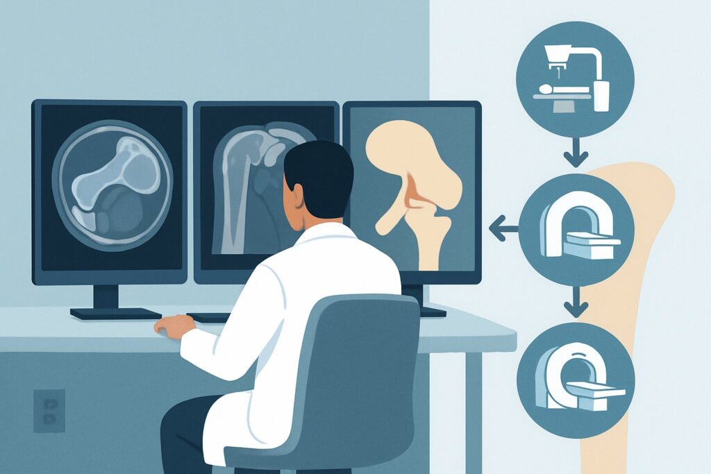
For evaluating suspected labral pathology or instability, the ACR designates both MR arthrography shoulder and MRI shoulder without IV contrast as Usually Appropriate. However, MR arthrography often provides a diagnostic advantage in this specific clinical context.
The procedure involves injecting a dilute gadolinium-based contrast agent directly into the glenohumeral joint space under fluoroscopic or ultrasound guidance. This distends the joint capsule, forcing contrast into subtle tears and separating structures that would otherwise be apposed. This technique significantly increases the sensitivity and specificity for detecting labral tears, especially small or non-displaced ones. It is also superior for evaluating the integrity of the glenohumeral ligaments and the joint capsule itself.
While a non-arthrographic MRI is also rated Usually Appropriate and avoids an injection, it relies on the presence of a native joint effusion to outline intra-articular structures. In a chronic setting without a significant effusion, a non-contrast MRI may fail to delineate a subtle labral tear, potentially leading to a false-negative result.
Why are other studies rated lower for this scenario?
- CT arthrography shoulder (May be appropriate): This study also uses intra-articular contrast to distend the joint and can be excellent for assessing the labrum and associated bony lesions (e.g., a bony Bankart lesion). It is a valuable alternative for patients with contraindications to MRI. However, it provides inferior soft-tissue contrast compared to MRI for evaluating the rotator cuff and surrounding muscles, and it involves significant ionizing radiation (adult_rrl=☢☢☢☢ 10-30 mSv).
- US shoulder (Usually not appropriate): Ultrasound is excellent for evaluating the rotator cuff tendons, biceps tendon, and subacromial-subdeltoid bursa. However, it cannot adequately visualize the deep, intra-articular structures like the labrum or the glenohumeral ligaments, making it the wrong test for the primary clinical question of instability or a labral tear.
Both MR arthrography and standard MRI have a radiation level of O (0 mSv). Given the superior detail for the suspected pathology, MR arthrography is often the preferred first choice when the clinical suspicion for a labral tear is high. Once you’ve decided on the best study for your patient, our protocol guide can help with the technical details. For a comprehensive overview of the non-contrast technique, see our guide: MRI Shoulder Without Contrast.
What’s Next After MR Arthrography? Downstream Workflow
The results of the MR arthrogram will guide your next steps, typically leading down one of three paths.
If the study is positive for a significant labral tear (e.g., unstable SLAP tear, Bankart lesion): The next step is a referral to an orthopedic surgeon. The imaging findings, correlated with the patient’s symptoms and functional demands, will determine whether surgical intervention (e.g., arthroscopic labral repair) is indicated. The detailed report from the MR arthrogram is crucial for surgical planning.
If the study is negative for a discrete structural lesion: A negative, high-quality MR arthrogram makes a significant labral tear unlikely. The focus should shift to a diagnosis of functional instability or another pain generator. The next step is typically a renewed emphasis on conservative management, including a structured physical therapy program focused on strengthening the dynamic stabilizers of the shoulder (rotator cuff and periscapular muscles). If pain persists despite therapy, you might reconsider the diagnosis, potentially looking at the sibling scenario for a suspected biceps tendon abnormality.
If the study is indeterminate or shows subtle findings: Sometimes, imaging may reveal labral fraying or degenerative changes without a clear, unstable tear. In these cases, the clinical correlation is paramount. An image-guided intra-articular anesthetic and/or corticosteroid injection can be both diagnostic and therapeutic. If the patient’s pain is temporarily relieved by the anesthetic, it confirms an intra-articular source. This may support a trial of conservative therapy or, in refractory cases, lead to consideration for diagnostic arthroscopy.
Pitfalls to Avoid (and When to Get Help)
Navigating the workup for suspected instability requires careful consideration to avoid common errors.
- Pitfall 1: Ordering a non-contrast MRI for high suspicion of a labral tear. While rated Usually Appropriate, a standard MRI can miss subtle tears if no joint effusion is present. If your clinical suspicion is high, the arthrogram provides higher diagnostic confidence.
- Pitfall 2: Not providing adequate clinical history. The radiologist’s ability to detect subtle pathology is enhanced by knowing the specific clinical concern (e.g., “Rule out posterior labral tear in a throwing athlete” is more helpful than “chronic shoulder pain”).
- Pitfall 3: Forgetting that instability is a dynamic problem. Static imaging like MRI captures a single moment in time. A patient with functional instability from poor neuromuscular control may have normal-appearing anatomy. Clinical exam remains essential.
If a patient has signs of a locked shoulder or a history of recurrent dislocations with persistent, severe apprehension, an earlier referral to orthopedics, even pending advanced imaging, is warranted.
Related ACR Topics and Tools
This article focuses on a single, specific clinical question. For a comprehensive overview of imaging for all common presentations of chronic shoulder pain, from rotator cuff disease to osteoarthritis, see our parent guide.
- For breadth across all scenarios in Chronic Shoulder Pain, see our parent guide: Chronic Shoulder Pain: ACR Appropriateness Decoded.
- To explore other clinical scenarios and their ACR-recommended imaging pathways, use the ACR Appropriateness Criteria Lookup.
- For detailed procedural techniques on recommended studies, browse the Imaging Protocol Library.
- To discuss radiation exposure from CT or other modalities with your patients, consult the Radiation Dose Calculator.
Frequently Asked Questions
Why is MR arthrography considered better than a standard MRI for a suspected labral tear?
MR arthrography involves injecting contrast directly into the joint, which distends the capsule and flows into any tears in the labrum or ligaments. This physical separation of tissues makes subtle or non-displaced tears much more conspicuous than on a standard (non-contrast) MRI, where structures may remain collapsed together, hiding the pathology. While a standard MRI is also highly rated, the arthrogram offers superior diagnostic confidence for this specific question.
What if my patient has a contrast allergy or refuses the injection for an arthrogram?
If a patient cannot undergo MR arthrography due to a gadolinium allergy, claustrophobia, or refusal of the injection, two main alternatives exist. A high-quality 3T non-contrast MRI of the shoulder is the best non-invasive option and is also rated ‘Usually Appropriate’ by the ACR. If MRI is contraindicated entirely (e.g., due to an incompatible implanted device), a CT arthrogram is a ‘May be appropriate’ alternative that provides excellent detail of the labrum and bone, though with less soft tissue detail and with ionizing radiation.
Is a CT arthrogram ever the first-choice study in this scenario?
Rarely. MR arthrography is generally preferred because of its superior ability to visualize soft tissues, including the rotator cuff muscles and tendons, without using ionizing radiation. However, a CT arthrogram might be considered first-line if there is a strong suspicion of an associated bony injury, such as a significant bony Bankart or Hill-Sachs lesion, as CT provides superior bone detail. It is also the best alternative for patients with absolute contraindications to MRI.
My patient’s MR arthrogram was negative, but their symptoms of instability persist. What now?
A negative high-quality MR arthrogram makes a significant structural cause of instability (like a major labral tear) unlikely. The focus should shift to ‘functional’ or ‘dynamic’ instability, which is often due to poor neuromuscular control and muscle patterning. The next step is typically a dedicated physical therapy program focusing on strengthening the rotator cuff and periscapular stabilizers. If symptoms remain severe and refractory to extensive conservative management, an orthopedic consultation for potential diagnostic arthroscopy might be considered.
Does the type of labral tear suspected (e.g., SLAP vs. Bankart) change the imaging recommendation?
No, the initial imaging recommendation of MR arthrography (or non-contrast MRI) remains the same regardless of the suspected location of the labral tear. Both SLAP (superior) and Bankart (anteroinferior) tears are intra-articular pathologies that are best visualized by distending the joint with contrast. The clinical history and physical exam findings that point toward a specific type of tear are critical information to include on the imaging requisition to help guide the radiologist’s search pattern.
Reviewed by Pouyan Golshani, MD, Interventional Radiologist — June 27, 2026