Which Imaging Study Is Best for Finger Joint Malalignment Without Fracture?
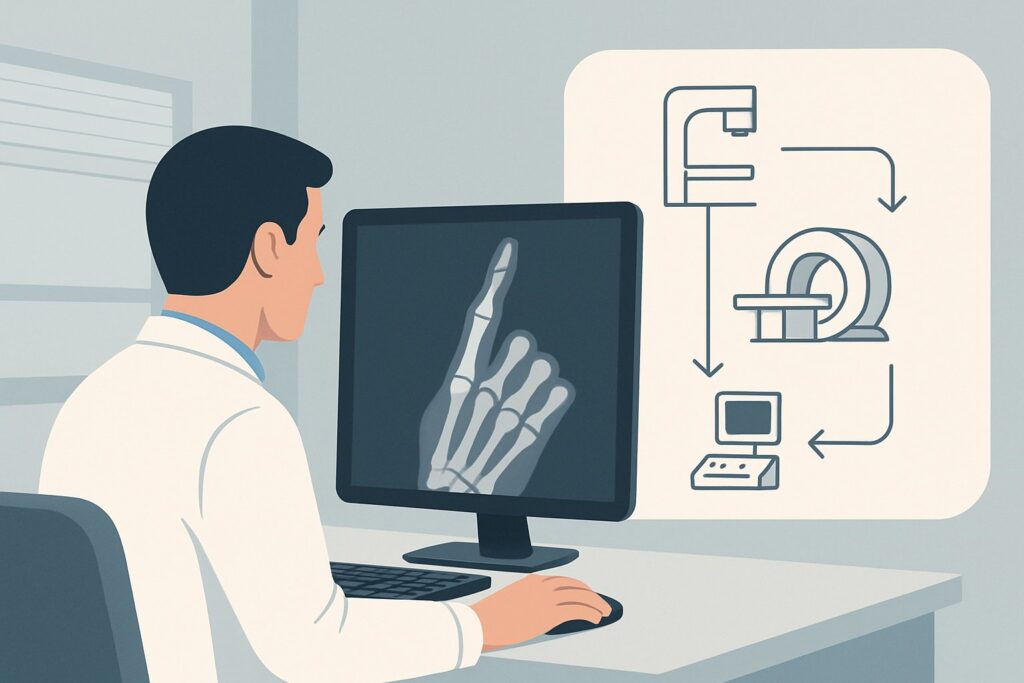
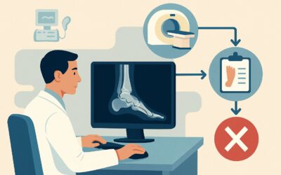
A 28-year-old patient presents to the urgent care clinic after jamming their finger during a weekend basketball game. They report immediate pain and deformity of the proximal interphalangeal (PIP) joint. On examination, the joint is swollen and tender. Initial radiographs confirm a PIP joint dislocation, which is successfully reduced, but you note persistent laxity on gentle stress testing. Crucially, the x-rays show no associated fracture. You are now faced with a clinical decision: is the joint stable, or is there a significant soft-tissue injury requiring surgical consultation? This article provides a step-by-step workflow for this exact scenario: determining the next imaging study when initial radiographs show metacarpophalangeal (MCP), proximal interphalangeal (PIP), or distal interphalangeal (DIP) joint malalignment in the absence of fracture. According to the American College of Radiology (ACR) Appropriateness Criteria, a US hand is a Usually Appropriate next step.
Who Fits This Clinical Scenario for Finger Joint Malalignment?
This guidance is specifically for patients who have undergone initial radiography for acute hand trauma and meet two key criteria:
1. Radiographs show malalignment: There is evidence of subluxation or dislocation at one of the finger joints (MCP, PIP, or DIP).
2. Radiographs show no fracture: The malalignment is an isolated finding without an associated osseous fracture.
This distinction is critical. The primary clinical question is not about a bone injury, but about the integrity of the soft-tissue stabilizers (ligaments, tendons, and the joint capsule) that are responsible for the joint’s instability.
It is equally important to know when this workflow does not apply. This guidance should be deferred if the patient presentation matches a different clinical variant:
- If a fracture is present: The scenario changes. The workup is now guided by the “Acute hand fracture on radiographs. Suspect hand tendon or ligament trauma. Next imaging study” variant, which has a different set of considerations.
- If malalignment is at the wrist: If radiographs show instability at the distal radioulnar joint (DRUJ) or between the carpal bones, that falls under the “Initial radiographs showing distal radioulnar joint or carpal malalignment” variant.
- If initial radiographs are normal: If the x-rays are entirely negative but you still have a high clinical suspicion for an occult injury (like a scaphoid fracture or ligament tear), you should consult the “Suspect acute hand or wrist trauma. Initial radiographs negative or equivocal” variant.
What Diagnoses Are You Working Up in This Scenario?
When a finger joint dislocates without a fracture, the injury is, by definition, to the soft tissues that provide stability. The purpose of further imaging is to identify and characterize these injuries to guide treatment, which can range from simple splinting to surgical repair.
The most common and significant diagnosis in this setting is a collateral ligament tear. Each finger joint is stabilized on its medial and lateral sides by these strong ligaments. A complete tear, especially of the ulnar collateral ligament of the thumb’s MCP joint (a “Skier’s thumb” or “Gamekeeper’s thumb”), can lead to chronic instability and arthritis if not properly managed. A Stener lesion, where the torn end of the ligament is displaced and trapped, will not heal without surgery and must be identified.
Another key consideration is a volar plate injury. The volar plate is a thick ligamentous structure on the palmar side of the PIP joint that prevents hyperextension. A significant tear or avulsion (which may be radiographically occult) can result in a “swan neck” deformity or persistent dorsal dislocation.
Less commonly, a tendon entrapment or subluxation can cause persistent malalignment. After a dislocation is reduced, a flexor or extensor tendon can become trapped within the joint space, preventing a stable, congruent reduction. At the MCP joint, an injury to the sagittal band of the extensor mechanism can cause the extensor tendon to subluxate, leading to functional deficits.
Why Is Ultrasound of the Hand the Recommended Next Study?
The ACR rates US hand as Usually Appropriate for this clinical scenario. The rationale is based on ultrasound’s unique ability to directly answer the primary clinical question: is the joint stable?
Ultrasound provides high-resolution imaging of the superficial soft tissues of the hand, including the very ligaments and tendons in the differential diagnosis. Its primary advantage is the ability to perform a dynamic assessment. The operator can apply gentle stress to the joint in real-time (e.g., valgus or varus stress for collateral ligaments) and directly visualize and measure any abnormal joint space widening or ligamentous disruption. This functional information is invaluable for determining stability and is not available with static imaging modalities. Ultrasound is highly effective for identifying full-thickness collateral ligament tears, Stener lesions, and significant volar plate injuries.
MRI hand without IV contrast is also rated Usually Appropriate. It offers excellent anatomic detail of all soft-tissue structures and is a valid alternative, particularly if ultrasound is unavailable or the findings are equivocal. However, MRI is a static examination, more costly, and less accessible than ultrasound. It cannot provide the real-time functional assessment that makes ultrasound so powerful in this context.
Other imaging studies are rated lower for specific reasons:
- CT hand (without or with contrast): Rated Usually not appropriate. While excellent for bone, CT provides poor soft-tissue contrast and cannot adequately visualize the small ligaments and tendons of the fingers. Since a fracture has already been excluded, CT adds little diagnostic value and exposes the patient to ionizing radiation (adult RRL ☢ <0.1 mSv).
- MRI hand with IV contrast: Rated Usually not appropriate. Intravenous contrast does not typically improve the visualization of acute traumatic ligament or tendon tears, adding unnecessary cost and potential risk.
What’s the Next Step After a Hand Ultrasound?
The results of the hand ultrasound will directly guide the subsequent clinical workflow, determining whether conservative management is sufficient or if a surgical consultation is necessary.
- If the study is positive for an unstable injury: A finding of a complete collateral ligament tear (especially with evidence of a Stener lesion), a significant volar plate rupture, or an entrapped tendon is an indication for urgent referral. The next step is a consultation with a hand surgeon to discuss operative repair.
- If the study is negative: A normal ultrasound demonstrating intact ligaments and tendons with no instability on dynamic stress testing is reassuring. This confirms that the joint is stable. The patient can typically be managed conservatively with a short period of immobilization (e.g., buddy taping or splinting) followed by physical therapy to restore range of motion.
- If the study is indeterminate: In cases where ultrasound findings are unclear due to significant patient swelling, pain, or other technical limitations, the next logical step is to proceed with the other Usually Appropriate study: MRI hand without IV contrast. MRI can provide definitive anatomic detail when dynamic assessment is not possible or inconclusive.
Pitfalls to Avoid (and When to Get Help)
Navigating this scenario requires attention to a few common pitfalls to ensure optimal patient outcomes.
First, avoid the pitfall of assuming stability after reduction. The absence of a fracture on x-ray does not mean the joint is stable. A thorough physical exam assessing for laxity is crucial, and a low threshold for advanced imaging is warranted if there is any doubt.
Second, do not delay imaging for a suspected unstable injury. Certain conditions, like a Stener lesion of the thumb UCL, have much better outcomes with early surgical repair. A delay can lead to ligament retraction and scarring, making a primary repair more difficult.
Third, ensure the ultrasound order is specific. Clearly state the clinical concern (e.g., “Rule out right 3rd PIP ulnar collateral ligament tear, assess for stability”) to ensure the sonographer performs the necessary dynamic stress maneuvers.
If you identify a complete ligament tear, a displaced intra-articular structure, or an irreducible dislocation, escalate immediately by consulting a hand surgeon.
Related ACR Topics and Tools
For a comprehensive overview of imaging for hand and wrist injuries, further reading and specialized tools can provide additional context.
For breadth across all scenarios in Acute Hand and Wrist Trauma, see our parent guide: Acute Hand and Wrist Trauma: ACR Appropriateness Decoded.
To explore other clinical situations or refine your imaging choices, you can use the following resources:
- ACR Appropriateness Criteria Lookup — for adjacent scenarios
- Imaging Protocol Library — for technique on the recommended study
- Radiation Dose Calculator — for cumulative dose conversations
Frequently Asked Questions
Why not just get an MRI for everyone with a finger dislocation?
While MRI is also rated ‘Usually Appropriate’ and provides excellent anatomic detail, ultrasound is often preferred as the initial study. Ultrasound’s key advantage is its ability to perform real-time dynamic stress testing, which directly assesses joint stability—the primary clinical question. It is also more accessible, faster, and less expensive than MRI.
What is a Stener lesion and why is it important to diagnose?
A Stener lesion occurs specifically with a tear of the ulnar collateral ligament (UCL) of the thumb’s MCP joint. The torn end of the ligament gets displaced superficial to the adductor aponeurosis, a nearby tendon sheath. This displacement prevents the ligament from healing back to the bone on its own. It is a clear indication for surgery, and missing this diagnosis can lead to chronic instability and arthritis of the thumb.
Is CT ever useful in this specific scenario of malalignment without fracture?
For this specific scenario, CT is rated ‘Usually not appropriate.’ Its strength is in defining bone anatomy and complex fractures. Since a fracture has already been ruled out by radiographs, and the clinical question is about soft-tissue integrity (ligaments, tendons), CT offers very little diagnostic information and exposes the patient to unnecessary radiation.
Can I just splint the finger and see how the patient does without further imaging?
This approach, known as a trial of conservative management, can be risky if a significant unstable injury is present. While a simple sprain may heal well with splinting, an undiagnosed complete ligament tear (like a Stener lesion) or a volar plate rupture will not heal properly and can result in permanent instability, stiffness, or deformity. Advanced imaging is recommended to avoid missing these surgically-correctable injuries.
Does this guidance apply to chronic or old finger dislocations?
No, this ACR guidance is specifically for acute trauma. The workup for chronic instability or a missed dislocation is different and often involves a different set of imaging considerations, where MRI or CT arthrography might play a larger role to assess for secondary arthritic changes and the quality of remaining soft tissues.
Reviewed by Pouyan Golshani, MD, Interventional Radiologist — May 29, 2026