

What Is the Best Imaging for Local Recurrence Surveillance of a Primary Bone Tumor?
ACR-guided imaging workflow for Malignant or aggressive primary bone tumor. Surveillance for local recurrence. (Malignant or Aggressive Primary Musculoskel
ACR-guided imaging workflow for Malignant or aggressive primary bone tumor. Surveillance for local recurrence. (Malignant or Aggressive Primary Musculoskel

ACR-guided imaging workflow for Malignant or aggressive primary musculoskeletal tumor with no suspected or known recurrence. Surveillance for pulmonary met
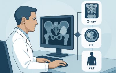
ACR-guided imaging workflow for Malignant or aggressive primary musculoskeletal tumor. Initial staging. Evaluation for extrapulmonary metastasis. (Malignan
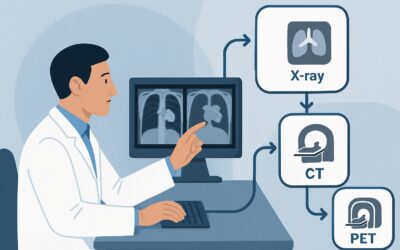
ACR-guided imaging workflow for Malignant or aggressive primary musculoskeletal tumor. Initial staging. Evaluation for pulmonary metastasis. (Malignant or

ACR-guided imaging workflow for Known osteonecrosis with articular collapse by radiographs. Surgery planned. Next imaging study. (Osteonecrosis): recommend
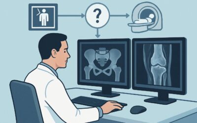
ACR-guided imaging workflow for Clinically suspected osteonecrosis. Normal radiographs or radiographs that show findings suspicious for osteonecrosis. Next

ACR-guided imaging workflow for Clinically suspected osteonecrosis. Initial imaging. (Osteonecrosis): recommended study, differential, downstream pathway.

ACR-guided imaging workflow for Previously healthy with worsening diffuse abdominal pain for 2 weeks. CTA shows occlusion of the superior mesenteric vein a

ACR-guided imaging workflow for Recent onset abdominal pain, peritoneal signs, and known atrial fibrillation. CTA shows filling defect in the proximal SMA

ACR-guided imaging workflow for Recent onset abdominal pain, no peritoneal signs, and known atrial fibrillation. CTA shows calcified atherosclerotic plaque