What Preoperative Imaging Is Best for Osteonecrosis with Articular Collapse?

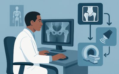
A 58-year-old man with a history of long-term corticosteroid use for an autoimmune condition presents to his orthopedic surgeon with progressively worsening left hip pain. Plain radiographs are definitive, showing advanced osteonecrosis of the femoral head with subchondral lucency and clear flattening of the articular surface, confirming collapse. A total hip arthroplasty is planned. To ensure optimal component sizing and placement, the surgeon needs to precisely delineate the extent of the necrotic bone and evaluate the remaining bone stock. This article details the ACR-guided imaging workflow for this specific preoperative scenario. According to the ACR Appropriateness Criteria, an MRI area of interest without IV contrast is Usually Appropriate for this indication.
Who Fits This Clinical Scenario for Preoperative Osteonecrosis Imaging?
This guidance is for a well-defined patient population where the diagnosis of osteonecrosis (ON) is already established and the disease has progressed to a critical stage.
Inclusion criteria for this workflow:
- A confirmed diagnosis of osteonecrosis from prior imaging, typically radiographs.
- Radiographic evidence of articular surface collapse (e.g., the “crescent sign,” subchondral fracture, or flattening of the femoral head).
- A surgical intervention, such as arthroplasty, core decompression with bone grafting, or osteotomy, is already planned.
This workflow is specifically for preoperative planning, not for initial diagnosis or for monitoring asymptomatic disease.
Exclusion criteria (patients who fit a different workflow):
- Initial Suspicion: If a patient presents with risk factors (e.g., steroid use, alcohol abuse, sickle cell disease) and joint pain but has not yet had imaging, they fit the “Clinically suspected osteonecrosis. Initial imaging” scenario.
- Suspicion with Normal Radiographs: If initial radiographs are negative or equivocal but clinical suspicion for ON remains high, that patient falls under the “Clinically suspected osteonecrosis. Normal radiographs…” scenario, which has a different imaging pathway.
This article focuses exclusively on the patient with known, collapsed ON who is on the path to the operating room.
What Are the Key Questions for Preoperative Imaging in Advanced Osteonecrosis?
In this scenario, the primary diagnosis is not in question. Instead, imaging is used to answer specific anatomical and pathological questions that directly influence surgical strategy and implant choice. The “differential” here is less about alternative diagnoses and more about characterizing the extent of disease and identifying potential surgical challenges.
Mapping the Extent of Necrotic Bone: The most critical question is defining the three-dimensional volume and location of the necrotic segment. Radiographs can show collapse but often underestimate the true extent of avascular bone. Precise mapping is essential for the surgeon to determine how much bone must be resected and to plan the placement and size of prosthetic components to ensure they are anchored in healthy, viable bone for long-term stability.
Assessing Articular Cartilage and Acetabular Integrity: While radiographs confirm bony collapse, they provide limited information about the overlying articular cartilage or the opposing articular surface (e.g., the acetabulum in the hip). Preoperative imaging can reveal the degree of cartilage delamination, identify chondral flaps or intra-articular loose bodies, and assess for secondary degenerative changes on the acetabular side, which may influence the choice between a hemiarthroplasty and a total joint replacement.
Evaluating Viability of Surrounding Bone: The success of joint-preserving procedures (less common in collapsed ON) and arthroplasty depends on the health of the surrounding bone. Imaging helps assess the extent of reactive bone marrow edema, which can correlate with the patient’s pain, and evaluates the structural integrity and viability of the bone that will support the implant. This is crucial for anticipating potential issues with implant fixation.
Why Is MRI Without Contrast the Recommended Preoperative Study for Collapsed Osteonecrosis?
For preoperative planning in a patient with known osteonecrosis and articular collapse, the American College of Radiology has determined that MRI area of interest without IV contrast is Usually Appropriate. This recommendation is based on its superior ability to answer the key surgical questions without unnecessary risks.
MRI provides unparalleled soft tissue and bone marrow contrast, making it the ideal modality for delineating the precise margins of the necrotic zone. T1-weighted sequences are highly sensitive for the characteristic low-signal intensity band that demarcates the necrotic segment from viable bone. Fluid-sensitive sequences (like T2-weighted or STIR) excel at identifying associated bone marrow edema, subchondral fluid, and the integrity of the articular cartilage. This detailed anatomical map allows the surgeon to plan resections and implant placement with high confidence.
In this specific scenario of established, collapsed ON, intravenous contrast adds little diagnostic value. The primary questions—the size of the necrotic segment and the extent of collapse—are answered by the morphological information on non-contrast sequences. Therefore, an MRI area of interest without and with IV contrast is rated as Usually Not Appropriate, as it adds cost and the small risk associated with gadolinium-based contrast agents without providing significant additional surgical planning information.
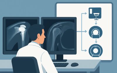
While CT area of interest without IV contrast is also rated Usually Appropriate, it is generally considered a secondary option to MRI. CT offers excellent visualization of bony architecture, such as the degree of femoral head flattening and the presence of subchondral fractures. It can be particularly useful if MRI is contraindicated (e.g., due to an incompatible implanted device) or if the surgeon’s primary concern is purely bone stock quantification. However, it lacks MRI’s ability to directly visualize bone marrow edema, cartilage health, and the viability of surrounding tissues, and it involves ionizing radiation (RRL=Varies).
Other modalities are rated Usually Not Appropriate. A Bone scan is non-specific and provides poor anatomical detail for surgical planning. MR arthrography is invasive and unnecessary when the primary goal is to assess bone, not subtle labral or ligamentous tears in isolation.
What’s Next After MRI? Downstream Workflow
The results of the preoperative MRI directly guide the final surgical plan and the conversation with the patient. The workflow is generally straightforward once this definitive imaging is complete.
- Findings Confirm Suitability for Planned Arthroplasty: In the vast majority of cases, the MRI will confirm the extent of necrosis and collapse seen on radiographs, providing the detailed anatomical map the surgeon needs. The findings on the size of the lesion, the quality of adjacent bone, and the status of the acetabulum will be used to select the appropriate type and size of prosthetic components. The next step is proceeding with the scheduled surgery.
- Findings Suggest a More Complex Procedure: Occasionally, the MRI may reveal that the extent of necrosis is far greater than anticipated, involving a significant portion of the femoral neck or acetabulum. This might necessitate a change in surgical plan, such as requiring a more complex implant with a longer stem, the use of bone grafts, or a custom component. The surgeon will use this information to ensure the correct equipment is available.
- Incidental or Unexpected Findings: Rarely, an MRI may reveal an unexpected finding, such as evidence of a low-grade infection or an underlying pathologic lesion. If findings are suspicious for infection, the surgical plan would be drastically altered to include aspiration for culture, and arthroplasty would be delayed. If a tumor is suspected, this would trigger a completely different diagnostic and management pathway, often involving an orthopedic oncologist.
In this preoperative setting, a “negative” or “indeterminate” result is highly unlikely, as the diagnosis and collapse are already known from radiographs. The MRI serves to characterize, not to diagnose.
Pitfalls to Avoid (and When to Get Help)
Even in this well-defined scenario, several pitfalls can complicate preoperative planning.
- Ordering Contrast Unnecessarily: Routinely adding IV contrast for preoperative ON assessment increases cost and exposes the patient to gadolinium without typically adding useful information for surgical planning. Stick to the non-contrast protocol unless a specific comorbidity (like suspected infection or tumor) warrants it.
- Ignoring the Opposing Joint Surface: Ensure the imaging field of view and the reporting radiologist adequately assess the opposing articular surface (e.g., the acetabulum). Unaddressed pathology there can lead to poor surgical outcomes.
- Relying on an Old MRI: If a significant amount of time has passed since the last MRI, and the patient’s symptoms have worsened, the degree of collapse and secondary arthritic change may have progressed. Consider repeating the study if the prior imaging is not recent enough to reflect the current anatomy.
- Misinterpreting Reactive Edema: Extensive bone marrow edema adjacent to the necrotic zone is a reactive change and should not be mistaken for infection or tumor, though in the right clinical context, these must be considered.
If the MRI report raises suspicion for a concurrent process like infection or malignancy, immediate escalation to or consultation with an orthopedic oncologist or infectious disease specialist is warranted before proceeding with arthroplasty.
Related ACR Topics and Tools
For a comprehensive overview of imaging recommendations across all clinical presentations of osteonecrosis, from initial suspicion to postoperative follow-up, please see our parent guide. It provides the breadth that complements this article’s depth.
- For breadth across all scenarios in Osteonecrosis, see our parent guide: Osteonecrosis: ACR Appropriateness Decoded.
- To explore other clinical scenarios, use the ACR Appropriateness Criteria Lookup.
- For details on imaging techniques, browse the Imaging Protocol Library.
- To discuss radiation exposure with patients, consult the Radiation Dose Calculator.
Frequently Asked Questions
Why is MRI without contrast preferred over CT when CT is also rated ‘Usually Appropriate’?
While CT provides excellent detail of the bone structure and collapse, MRI offers superior contrast resolution for assessing bone marrow viability, delineating the exact size of the necrotic lesion, evaluating cartilage health, and identifying reactive edema. This information is generally more valuable for comprehensive surgical planning. CT is a strong alternative if MRI is contraindicated.
Is there any role for intravenous contrast in this specific scenario?
For routine preoperative planning of known, collapsed osteonecrosis, IV contrast is rated ‘Usually Not Appropriate’ because it rarely adds information that changes the surgical plan. The key anatomical questions are answered with non-contrast sequences. Contrast would only be considered if there is a specific, superimposed concern, such as a suspected abscess or tumor, which falls outside this standard clinical scenario.
My patient has a pacemaker and cannot get an MRI. What is the next best step?
If a patient has a contraindication to MRI, a non-contrast CT of the area of interest is the recommended alternative. It is also rated ‘Usually Appropriate’ by the ACR for this scenario and will provide excellent detail on the bony collapse, subchondral fractures, and bone stock needed for surgical planning.
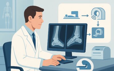
Does this guidance apply to all joints affected by osteonecrosis?
Yes, the principles of this guidance apply to any joint with advanced, collapsed osteonecrosis planned for surgery, including the hip, knee, shoulder, or ankle. The goal of preoperative imaging remains the same: to define the extent of necrosis and assess the integrity of the surrounding structures to plan the surgical intervention.
If the surgery is a joint-preserving procedure instead of arthroplasty, does the imaging recommendation change?
No, the recommendation for a non-contrast MRI remains the same. In fact, for joint-preserving procedures like core decompression with bone grafting, the information from an MRI is even more critical. The surgeon must have a precise map of the necrotic and viable bone to ensure the graft is placed effectively and the procedure has the best chance of success.
Reviewed by Pouyan Golshani, MD, Interventional Radiologist — May 30, 2026